Auditory II Central Auditory System
Auditory II Central Auditory System . I. Tonotopic organization (i.e. frequency map in the central auditory system) - Sound is represented tonotopically in the cochlea, which results in tonotopy in the auditory nerve fibers.

Auditory II Central Auditory System
E N D
Presentation Transcript
Auditory II Central Auditory System
I. Tonotopic organization (i.e. frequency map in the central auditory system) - Sound is represented tonotopically in the cochlea, which results in tonotopy in the auditory nerve fibers. - Tonotopy is preserved throughout central auditory system, e.g. as evident in a Golgi stain of the cochlear nuclei.
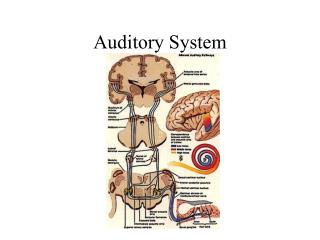
II. Neuroanatomy of the auditory system - Sequence (1) Auditory nerve fibers from cochlea terminate in cochlear nuclei (DCN or VCN) (2) From the CN, most neurons project to superior olivary nuclei for sound localization (3) The lateral lemniscus is a collateral path to the inferior colliculus.
(4) All ascending neurons (from SO and LL) terminate in the inferior colliculus (5) IC projects to the medial geniculate (thalamus) - Most commisures occur at the trapezoid body, and above the SO most neurons are bi-aural.
III. Sound localization by azimuth - Locating a sound source carries the additional problems of (1) distinguishing multiple sound sources over background noise and (2) suppressing the effect of echoes - Due to path length difference, a sound source will have different effects on the right and left ears.
(1) time difference: ITD and IPD; IPD is better perceived due to phase-locking of ANFs (2) loudness difference: ILD - Relative importance of ITD and ILD cues depends on stimulus frequency - The superior olivary nuclei compute sound localization. - Bushy cells have two major specializations to deal with very small ITDs: (1) do not display temporal summation and (2) very tight synapses with ANF
- MSO neurons measure the ITD - MSOs are activated by coincident stimulation by both ears - The ITD to which a MSO is most sensitive depends on the internal neural delay (function of path length of bushy cells) - LSO neurons measure the ILD - LSO neurons are stimulated when ipsilateral intensity is greater than contralateralintensity.
IV. Sound localization by elevation - It is more difficult to determine elevation of cue; “cone of confusion” is apparent in single tone stimulus - Sound reflections off the pinna differ depending on elevation of sound source; the resulting interference causes nulls at high frequency, and the location of the null is a function of elevation
V. Auditory (cortex) neurons may encode stimuli through coordinated firing in response to a stimulus VI. Descending systems - Nerves to the middle ear muscles dampen effect of loud sounds and help filter out low-frequency self-generated sounds (e.g. due to chewing) - Projections from the olivocochlear bundle aid hearing in noisy backgrounds.