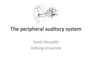
The auditory system
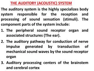
The auditory system. Structure and function. Objective. Recap – outer, middle and inner ear to BM response To continue the hearing process from the inner ear to the brain How the physical properties of sound, such as frequency and amplitude, are represented in the auditory system.

The auditory system
E N D
Presentation Transcript
The auditory system Structure and function
Objective • Recap – outer, middle and inner ear to BM response • To continue the hearing process from the inner ear to the brain • How the physical properties of sound, such as frequency and amplitude, are represented in the auditory system
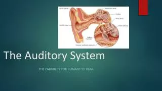
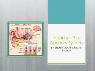
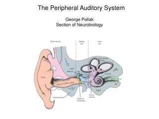
Organ of Corti • The organ of corti is the hearing sense organ and lies on the BM • It consists of supporting cells and hair cells • 2 groups of hair cells: inner and outer hair cells • Protruding from each hair cell are hairs called stereocilia • The tectorial membrane lies above the stereocilia –shearing motion between BM and tectorial membrane – causes stereocilia to be displaced
Inner hair cells • Function is to convert BM mechanical movement into neural activity – achieved in the following way (Moore, 2004): • The stereocilia are joined by fine links called ‘tip links’. • Deflection of the stereocilia leads to the opening of “transduction channels”. • Flow of potassium ions into the hair cell – voltage difference between the inside and outside of the hair cell.
Inner hair cells • Causes the release of neurotransmitter and the initiation of action potentials in the neurons of the audtiory nerve. • Action potential – ‘firing’ of a neuron. Propagation is in one direction only down the length of the axon • Most of the afferent neurons make contact with the inner hair cells • Possibly all information about the input sound is conveyed via the inner hair cells
Outer hair cells • Have a role in achieving high sensitivity and sharp tuning • Most of the efferent neurons synapse directly with the outer hair cells • Efferent neurons carry information from higher auditory system to cochlea • Afferent neurons carry information from the cochlea to the higher auditory system
Auditory nerve • Consists of vestibular and cochlear nerve • Cochlear nerve – the axon fibres of neurons whose cell bodies are in the spiral ganglion of the cochlea • dendrites of these neurons synapse with the hair cells – dendrites information receivers
Afferent auditory nerve • Transmits hearing information from cochlea to central nervous system • Important findings from recording impulses in single auditory nerve fibres. • Spontaneous firing, frequency selectivity of fibres, phase locking
Spontaneous firing rates • Firing in the absence of sound stimulation • Fibres divided into three groups based on their spontaneous firing rates • High, medium and low spontaneous rate fibres • High spontaneous rate fibres generally have lower thresholds and smaller dynamic ranges than medium or low spontaneous rate fibres
Frequency selectivity • Fibres show frequency selectivity – as fibres responding to activity at restricted regions of the BM • Characteristic frequency (CF) – frequency at which the threshold of a fibre is lowest • Fibres with high CF found in periphery of auditory nerve – orderly decrease in CF towards centre of the auditory nerve • Tonotopic organisation – place representation of frequency on BM maintained in auditory nerve
Phase locking • The firing of a neuron at one distinct point (phase) in the period of a sound wave. • temporal regularity in the firing pattern of a neuron in response to a periodic stimulus • upper limit: 4-5 kHz • May not fire on every cycle, but will occur at roughly the same phase of the waveform • Time intervals between spikes are approximately integer multiples of the period of the waveform • an indicator of the period of the waveform
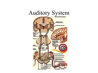
The brainstem and auditory cortex • Binaural perceptions, such as our ability to localise sounds depends on the interaction of information from both ears • The information from both auditory nerves is first combined in the brainstem • Highly complex system, many of the neural pathways within and between the nuclei have yet to be investigated • Brainstem nuclei: cochlear nucleus, superior olivary nucleus, inferior colliculus, medial geniculate body.
Tonotopic organisation of frequency is preserved – neurons aligned respective to the frequencies to which they are most sensitive • The responses of neurons at higher levels in the auditory system – not as well studied as responses in the auditory nerve • Some neurons in the auditory cortex only respond to complex stimuli, or to stimuli with time varying characteristics
Cochlear nucleus • The auditory nerve carries signals from the cochlea to the cochlear nucleus in the brainstem. • First brainstem nucleus at which afferent auditory nerve fibres synapse • Monaural – input from same side ear only • 3 parts – anteroventral, posteroventral, dorsal • Not clear what its function in auditory processing is. – tonotopic organisation of frequency
Superior olivary complex • Binaural - Here the inputs from both ears converge • Important for sound localisation • Use of timing and intensity differences
Inferior colliculus • Receives inputs from the olivary complex and the cochlear nucleus • Some cells are monaural, some are binaural • Much (but not all) of the input to the inferior colliculus comes from the opposite ear • May function in sound localisation, and in combining information from lower areas in the brainstem
Medial geniculate nucleus • Last stop in the auditory pathway before the cerebral cortex. • 2-way information flow between medial geniculate and the auditory cortex • Feedback from the brain is tightly integrated with sensory information flowing up to the brain
Auditory cortex • Primary auditory cortex (A1) – the first area within the temporal lobes of the brain responsible for processing acoustic information • Each cochlea has input to each auditory cortex • Tonotopic organisation of frequency is maintained in the A1 • 2-way information between cortex and brainstem
No response to steady, unchanging tones – pure tones • Some cortical neurons respond to tones increasing or decreasing in frequency • Some respond to amplitude variations
Next: frequency resolution (selectivity) and pitch perception, reading material