The Auditory System
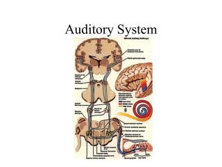
This overview highlights the key components of the auditory system, detailing the roles of perilymph and endolymph within the cochlea, and the structure of the membranous labyrinth. It describes the Organ of Corti, auditory ganglion, and cochlear nuclei, emphasizing their significance in sound localization and auditory processing. Key neural pathways in the brainstem, including the superior olivary complex and inferior colliculus, are covered to illustrate how auditory information is integrated and sent to the auditory cortex. Understanding these elements is crucial for comprehending auditory perception.

The Auditory System
E N D
Presentation Transcript
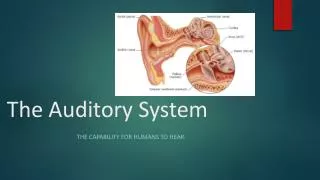
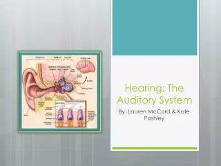
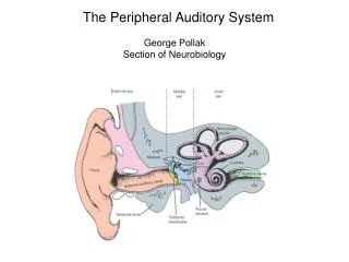
• Perilymph – resembles CSF, within bony labyrinth [low K, high Na] • Endolymph – within membranous labyrinth [high K, low Na] • Membranous labyrinth o Cochlear duct (scala media), scala vestibule, scala tympani (lower)
• Scalavestibuli and scala tympani communicate at the apex of cochlea via helicotrema • Organ of Corti – receptor organ of cochlea. It rests on basilar membrane o Single row of IHC o Band of OHC o Stereocilia project to tectorial membrane
o Apical surface of HC is bathed in endolymph, and basal surface in perilymph • Auditory (spiral) ganglion – in modiolus (bony core of cochlea) o Peripheral process – innervate IHC o Central process – form auditory branch of VIII
• Cochlear nuclei are lateral to inferior cerebral peduncles o VIII ascending branch to anteroventral cochlear nucleus o VIII descending branch to posteroventral CN and dorsal CN • Basilar membrane – high frequencies at base, low frequencies at apex.
o Three complete tonotopic representations of BM in dorsal and ventral cochlear nuclei • Localization of sound – important in auditory system.
Hence there are frequent decussations of axons across midline and terminations arising in both hemispheres in each of several nuclei to compare input from both ears.
o Only large lesions along the auditory path produces deficits in sound localization and none past the CN eliminates input from one ear • BS nuclei o Superior olivarycomplex – MSO and LSO are dorsolateral to medial lemniscus
• Medial superior olive – 1st site in CNS that receives inputs from two ears by way of ipsi and contra CN o Inferior colliculus – caudal part of midbrain tectum • Obligatory site of termination for almost all ascending auditory fibers
• BS tracts o Trapezoid body – long myelinated bundles crossing midline ventral to medial lemniscus • Axons of CN that cross midline to innervate contra SOC.
o Lateral lemniscus – myelinated fiber dorsolateral to medial lemniscus, and appears to grasp inferior colliculus.
• All axons from all lower BS auditory nuclei and carries them to IC o Brachium of IC – thin fiber tract that forms bump on dorsal surface just lateral to SC • These are axons IC MGN of thalamus • Medial geniculate complex – ventral to pulvinar
• MGN internal capsule temporal lobe • Auditory cortex – junction of central sulcus and lateral (Sylvian) fissure (lower bank) o Called superior temporal plane or “Heschl’sgyri) o Heavily myelinated area