THE AUDITORY (ACOUSTIC) SYSTEM
THE AUDITORY (ACOUSTIC) SYSTEM The auditory system is the highly specializes body system responsible for the reception and processing of sound sensation (stimuli). The component parts of the system include: The peripheral sound receptor organ and associated structures (The ear).

THE AUDITORY (ACOUSTIC) SYSTEM
E N D
Presentation Transcript
THE AUDITORY (ACOUSTIC) SYSTEM The auditory system is the highly specializes body system responsible for the reception and processing of sound sensation (stimuli). The component parts of the system include: The peripheral sound receptor organ and associated structures (The ear). The auditory pathway for conduction of nerve impulse generated by transduction of mechanical sound waves by the sound receptor organ Auditory processing centers of the brainstem and cerebral cortex
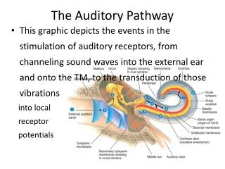
THE EAR • The ear consists of the auditory apparatus and the organs concerned with maintenance of balance. • It is subdivided into three parts viz. • The external ear, • The middle ear and • The inner (Internal) ear (Fig 1).
THE EXTERNAL EAR The external ear consists of • The pinna or auricle, which is responsible for the collection of sound waves and • The external acoustic meatus, which transmits the sound waves to the tympanic membrane at the lateral wall of the middle ear. • The pinna consists mainly of a thin plate of yellow elastic fibrocartilagineous framework to which is firmly attached a thin skin. • The external features of the pinna are illustrated in Fig 2
The external acoustic meatus measures about 2.5cm long and runs a peculiar S-shaped sinuous course towards the tympanic membrane at the lateral wall of middle ear. • The canal is covered by skin which is adherent to the wall and contains • hair, • sebaceous and • Numerous ceruminuous glands. • These appendages prevent the entry of particulate body into the canal.
The tympanic membrane (Eardrum) • This lies between the meatus and the middle ear (Tympanic cavity). • It is a thin, disc-shaped fibrous sheet, lined internally by mucous membrane and externally by skin The Tympanic Cavity (Middle Ear) • This is the slit-like cavity in the pitrous part of the temporal bone. • It accommodates the acoustic ossicles, which are responsible for the conduction of sound waves through the cavity. • The cavity is disc-shaped in transverse section and comprises six walls viz. posterior, anterior, lateral, medial, inferior and superior.
The ossicles are connected in the order, Malleus, Incus and Stapes. The malleus is attached to the tympanic membrane of the lateral wall, while the stapes is attached to the oval window of the medial wall. • The middle ear cavity also contains two muscles, tensor tympany and stapedeus which control the vibrations of the ossicles. • It also communicates anteriorly with the nasopharynx and posteriorly with the mastoid air cells • Four nerves are also located in the cavity. These are: • Chorda tympani nerve • Tympanic nerve • The tympanic plexus, and • The lesser petrosal nerve.
THE INTERNAL EAR • This part is also located in the pitrous part of the temporal bone and consists essentially of a complicated bony labyrinth, which is made up of a central part called the vestibule. • The vestibule communicates posteriorly with three semicircular canals, and anteriorly with the coiled, shell-shaped cochlear. • Within the entire bony labyrinth is the membranous labyrinth surrounded by perilymph. • The membranous labyrinth adopts the shape of the bony labyrinth, contains endolymph and consists of three parts viz.
Two small communicating sacs, the utricle and the saccule, both located in the vestibule. • Three semicircular ducts located in the semicircular canals. • The cochlear duct, which is located in the cochlear. • The diagram below illustrates the external features of the bony and membranous labyrinths.
There are specialized sensory receptor areas in each component of the membranous labyrinth. These are: • The maculae of the utricle and saccule, which record the direction of gravitational field relative to the head (Vestibulation/Balancing) • The cristaampullaris of the semicircular canals, which records movements in the endolymph resulting from rotational movements of the head (Vestibulation/Balancing). • The organ of corti (Spiral organ) of the cochlear duct, which respond to vibrations induced in the endolymph by sound waves transmitted by the stapes at the oval window (Hearing).
THE AUDITORY PATHWAY • The first order neurons (1st order) of the auditory pathway are: • The spiral ganglion cells of the organ of Corti. Most of the central processes of these cells are myelinated. • These processes form the cochlear nerve, which traverses the internal acoustic meatus alongside vestibular and facial nerve fibers. • Cochlear fibers accompanied by vestibular fibers pass to the lateral aspect of the brainstem at the junction of the pons and medulla oblongata. • Some of the cochlear fibers terminate in the ventral cochlear nucleus (2nd order) while others terminate in the dorsal cochlear nucleus (2nd order).
Axons of neurons in the ventral and dorsal cochlear nuclei cross the midline at the caudal aspect of the pons. • These crossing fibers constitute the Trapezoid body. • After crossing, trapezoid fibers turn upwards. Some of these ascending fibers (From the ventral cochlear nucleus) terminate in the superior olivary nucleus (3rd order) while some others (From both nuclei) continue beyond this nucleus and are joined by fibers from the superior olivary nucleus to constitute the lateral lemniscus.
Fibers of the lateral lemniscus proceed to the inferior colliculus where they synapse with Neurons of the inferior colliculus (4thorder Neurons). • Collicular neurons give rise to fibers, which proceed to the medial geniculate body of the thalamus. • Neurons of the medial geniculate body (5th order) give rise to fibers, which constitute the auditory radiations. • These fibers project to the inferior temporal gyrus where they terminate in • The primary auditory area (Brodmann’s area 41 & 42).