Auditory (Cochlear) System
Auditory (Cochlear) System. II. How Sound is Transduced into Electrical Events. Auditory apparatus composed of : external, middle and internal ear. Tympanic membrane vibrates on receiving sound waves.

Auditory (Cochlear) System
E N D
Presentation Transcript
II. How Sound is Transduced into Electrical Events • Auditory apparatus composed of : external, middle and internal ear. • Tympanic membrane vibrates on receiving sound waves. • Ossicles of middle ear transmit vibrations of tympanic membrane to foot plate of stapes which is attached to oval window. • Movement of stapes causes pressure in the perilymph of scala vestibuli and displacement of basilar membrane to which the Organ of Corti (containing the hair cells) is attached. • Movement of basilar membrane causes the hairs of hair cells to sheer back and forth in the tectorial membrane. • Sheering movements of the hairs generates a depolarizing (receptor) potential in the hair cells and transmitter to be released onto the peripheral terminals of cochlear nerve fibers (cell bodies in the spiral ganglion). • Summation of synaptic potentials generates an action potential in the cochlear nerve fiber
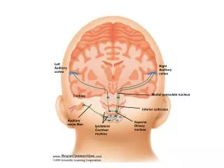
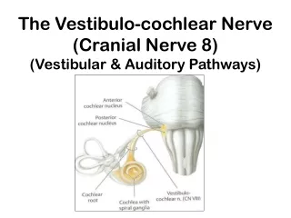
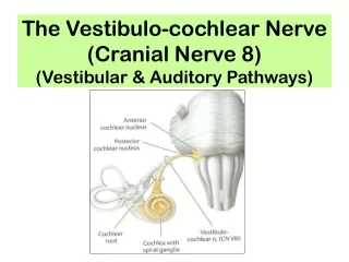
III. Major Nuclear centers and Pathways1st Order Neuron • Cell body localized in the spiral ganglion of the cochlear modiolus. • Peripheral process receives input from the hair cells (hair cell is presynaptic to terminals of process). • Central processes of ganglion cells form the cochlear nerve (part of VIII cranial nerve) which enters the brainstem at the cerebellopontine angle. • Root fibers of the cochlear nerve terminate on 2nd order neurons in the cochlear nuclei (dorsal and ventral) located at the level of the rostral medulla.
III. Centers and Pathways (Cont.)2nd Order Neuron • Cell body localized in the dorsal and ventral cochlear nuclei. All the fibers of the cochlear nerve terminate in the cochlear nuclei. • Axons cross the midline in the dorsal, intermediate and ventral (trapezoid body) acustic striae and join the ascending auditory pathway (aka lateral lemniscus). • Some axons of the trapezoid body end bilaterally in the superior olivary nuclear complex and nuclei of the trapezoid body (both these centers contain 3rd order neurons). • Unilateral lesions in the auditory nerve or cochlear nuclei (both dorsal and ventral) cause complete deafness of the ipsilateral ear.
III. Centers and Pathways (Cont.)3rd Order Neuron and lateral lemniscus • Cell bodies in the sup olivary nu complex or nu of trapezoid body. • Axons ascend ipsi- and contralaterally and join the ascending auditory pathway (lateral lemniscus). • Because each lateral lemniscus (LL) above the medulla has crossed and uncrossed fibers (inputs from both ears), unilateral lesions of the LL will result in bilateral hearing deficits that are more pronounced on the ear contralateral to the lesion. • All the fibers of the LL terminate in the inferior colliculus (part of the tectum at caudal midbrain levels).
III. Centers and Pathways (Cont.)4th and Higher Order Neurons • Neurons in the inferior colliculus communicate with each other by way of the commissure of the inf colliculus. • Axons of inf colliculus neurons ascend in the brachium of the inf colliculus to the ipsilateral medial geniculate nucleus, part of the thalamus. All the axons originating in the inf colliculus terminate in the med geniculate nucleus. • Cells in the med geniculate send their axons by way of the auditory radiation to the transverse temporal gyri of Heschl in the temporal lobe. • Ant transverse gyrus is the primary auditory area (Brodman’s area 41). • Post transverse gyrus is the auditory association area (area 42). • Unilateral lesions in the inf colliculus, med geniculate nucleus, auditory radiation or gyri of Heschl cause bilateral hearing loses that are more prominent on the ear opposite the lesion
IV. Clinical Correlation • Conduction deafness related to passage of sound waves through external or middle ears. • Perception deafness attributed to pathology of hair cells, cochlear nerve. • Central deafness is due to lesions of the auditory pathway in the CNS. • Presbycusis is the gradual hearing loss with aging. • Auditory Evoked Pontials (AEP) are used to pinpoint where lesion along the auditory pathway might occur. Peak I is generated by cochlear nerve, Peak II occurs at or near cochlear nuclei, Peak III is generated at the level of the lower pons in the region of sup olivary complex and trapezoid body, Peak IV and V occur at level of upper pons or lower midbrain which correspond to the location of lateral lemniscus and inferior colliculus.