Auditory Brainstem
Auditory Brainstem. Cochlear Nucleus. Auditory Brainstem . Formally includes Cochlear nuclei (medulla) Superior Olivary Complex (pons) Nuclei of Lateral Lemniscus (midbrain). Cochlear Nucleus . Each ANF innervates all three CN divisions: Anteroventral CN (AVCN) Posteroventral CN (PVCN)

Auditory Brainstem
E N D
Presentation Transcript
Auditory Brainstem Cochlear Nucleus
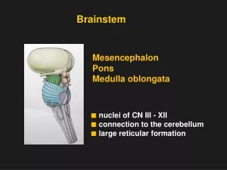
Auditory Brainstem • Formally includes • Cochlear nuclei (medulla) • Superior Olivary Complex (pons) • Nuclei of Lateral Lemniscus (midbrain)
Cochlear Nucleus • Each ANF innervates all three CN divisions: • Anteroventral CN (AVCN) • Posteroventral CN (PVCN) • Dorsal CN (DCN) AVCN DCN ANFs PVCN
Cochlear Nucleus: “Cochleotopic” (tonotopic) Organization • Each ANF innervating an IHC receptor projects to second order neurons in each division of CN within a single 2D plane (or lamina). • Each isofrequencylamina processes information from a single point along the basilar membrane (“cochlear channel”). Osen (1969) Lorente de No (1981)
Cochlear Nucleus: “Cochleotopic” (tonotopic) Organization • Each CN division has a complete, topographically organized, “cochleotopic” projection of the Organ of Corti. • Cochleotopic organization gives rise to “tonotopic” gradient (a “map”) of sound frequency along a particular axis within each nucleus. Tonotopic gradient in DCN (Rose 1960)
Cell Types found in VCN • CN contains at least 9 morphologically distinct cell types. • VCN contains • Spherical bushy cells (rostral AVCN) • Globular bushy cells (caudal AVCN) • Stellate/multipolar cells (AVCN/PVCN) • Octopus cells (PVCN) • Granule cells (similar to cerebellum).
Cells found in DCN Cells in DCN • Five cell types: • Granule • Fusiform • Giant • Cartwheel • Stellate Lorente de No (1981)
Inhibition first arises in CN Wickesburg & Oertel • Interneurons (mostly multipolar and granule cells) make intrinsic inhibitory connections between neurons within (local) and among the three divisions • Tuberculoventral loop: • Provides focal inhibitory feedback from DCN to VCN neurons. • Feedback inhibition may suppress echoes in reverberant environments. GABA GLY
CN: Functional organization and neuronal properties • Stimulus features (spectrum, intensity, time course, etc.) are differentially encoded by the different morphological cell types in the CN. • Each cell type targets specific higher nuclei for parallel processing of sound components.
Transformation of Discharge Patterns in the CN • Discharge patterns show much more complexity than ANFs. • Both tonic (sustained) and phasic (brief) responses occur to CF tones • Many discharge patterns are shaped by time-dependent interactions of excitation and inhibition.
Transformation of Spectral Receptive Field • Response areas, particularly in the DCN, become more elaborate with both excitatory and inhibitory regions. • Five classes of spectral response areas are found in CN.
Type I Response Area • Simple excitatory response area (no inhibitory areas). • Resembles ANF tuning properties. • Associated with large complex synaptic endings (“end bulbs of Held”) in VCN.
Type III Response Area • Excitatory response area like Type I, but with one or more flanking inhibitory areas (lateral inhibition). • Tonal response inhibited by broadband background noise. • Expressed in VCN and DCN choppers, DCN pausers.
Type IV Response Area • Very complex: One or more excitatory response areas, widespread inhibitory response areas at higher sound levels. • Mainly associated with DCN fusiform cells. • Show variety of discharge patterns (pauser, buildup, chopper, onset) depending upon stimulus frequency.
Type IV Responses to Tones and Noise • Response of Type IV unit can be excitatory or inhibitory, depending upon the tone frequency and level. • Type IV units integrate over wide frequency range.
Inhibition Shapes Response Properties in CN • Inhibitory feedback alters the temporal features of signal transmission between AN and CN neurons. Kopp-Scheinpflug et al. (2002)
Inhibition Shapes Response Properties in CN • Inhibitory feedback alters the spectral features of signal transmission between AN and CN neurons. Kopp-Scheinpflug et al. (2002)
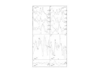
Temporal Features are Enhanced by CN Circuitry • Amplitude Modulation • ANFs phase-lock to AM envelopes only at low sound levels. Frisina et al. (1990)
Temporal Features are Enhanced by CN Circuitry • Amplitude Modulation • CN neurons enhance temporal coding of AM envelopes. • Best enhancement seen in neurons showing onset or chopper discharge patterns for unmodulated tones. Frisina et al. (1990)
Temporal Features are Enhanced by CN Circuitry • Phase-locking • VCN Bushy cells show improved precision of phase locking relative to their ANF inputs. • Improvement attributed to coincidence detection. • Onset coding • Reduced latency jitter in VCN octopus cells • Also attributed to coincidence detection. Joris, et al. (1998)
Anatomical Segregation of Cochlear Nucleus Outputs • Three main output pathways: • Dorsal acoustic stria: DCN to contralateral midbrain (NLL, IC) • Intermediate AS of Held: PVCN to SOC (periolivary) • Ventral AS, or Trapezoid Body: AVCN to SOC bilaterally, contra midbrain • All project to VNLL and IC
Functional Segregation of CN Projections: Parallel Processing • Two partially-overlapping but largely segregated pathways can be defined: • Monaural, or “what", pathway, in which non-spatial information (spectral composition, temporal contrast, intensity, etc.) is processed. • Binaural, or “where" pathway, in which the spatial information from the two ears converges for sound localization.
Partial Segregation of Information Processing • Despite the apparent dichotomy of these two processing pathways, the same types of acoustic cues may be important for the analyses that occur in each. • E.g., spectral information is used in the "where" pathway for determining a sound's elevation; temporal information used for pitch perception in the "what" pathway is also used in the "where" pathway for determining a sound's azimuth (horizontal location).
The "What" Pathway: Neural Bases of pitch, loudness, timbre perception. • "Monaural" pathway • Spectral and temporal features of the stimulus are processed and classified with minimal regard to location in space. • Arises from components of all three divisions of the cochlear nucleus, with a heavy emphasis on the DCN.