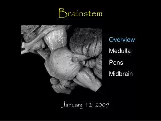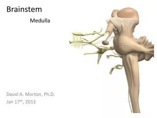
Brainstem
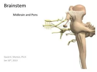
Midbrain and Pons. Brainstem. David A. Morton, Ph.D. Jan 10 th , 2013. Objectives. Explain how spinal nerves differ from cranial nerves Name all the cranial nerves and know their components and functions Identify and locate the CN’s associated with the medulla, pons and midbrain

Brainstem
E N D
Presentation Transcript
Midbrain and Pons Brainstem David A. Morton, Ph.D. Jan 10th, 2013
Objectives • Explain how spinal nerves differ from cranial nerves • Name all the cranial nerves and know their components and functions • Identify and locate the CN’s associated with the medulla, pons and midbrain • Recognize the major internal and external landmarks on the dorsal and ventral surface of the brain stem, so that you can determine if a gross or stained cross section is medulla, pons or midbrain. • Identify on a typical cross section all the brain stem nuclei containing motor neurons that end on striated muscle. • List the cranial nerves that contain parasympathetic fibers, the location of their nuclei, and their function • Explain why cranial nerves are so important in localizing lesions. • Name reflexes that test these nerves and brain stem levels. • Relate branches of the vertebrobasilar blood supply to the medulla and pons explaining the deficits that would occur with vascular occlusion.
Cranial nerve overview • CN I – Olfactory nerve
Cranial nerve overview • CN II – Optic nerve
Cranial nerve overview • CN III – Oculomotor nerve
Cranial nerve overview • CN IV – Trochlear nerve
Cranial nerve overview • CN V – Trigeminal nerve
Cranial nerve overview • CN VI – Abducens nerve
Cranial nerve overview • CN VII – Facial nerve
Cranial nerve overview • CN VIII – Vestibulocochlear nerve
Cranial nerve overview • CN IX – Glossopharyngeal nerve
Cranial nerve overview • CN X – Vagus nerve
Cranial nerve overview • CN XI – Spinal accessory nerve
Cranial nerve overview • CN XII – Hypoglossal nerve
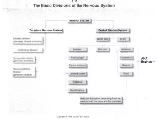
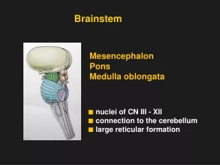
Brain Overview • Brainstem • Midbrain • Pons • Medulla Midbrain Pons Medulla
Brain Overview • Diencephalon • Thalamus • Hypothalamus • Pineal gland Th P HTh
Brain Overview • Corpus callosum • Lateral ventricle • 3rdventricle • Cerebral aqueduct • 4th ventricle Corpus callosum LV 3 aq 4
Spinal Cord and Spinal Nerve Review • Spinal nerve anatomy • Dorsal root • Somatic sensory neurons • Visceral sensory neurons • Ventral root • Visceral motor neurons • Somatic motor neurons Som S Alar VS Sulcus limitans VM SM Basal Spinal cord
Internal anatomy of brainstem • The fate of the alar and basal laminae • Why are brain stem sensory nuclei lateral to motor nuclei in brainstem? Som S Som S Alar Alar VS VS Sulcus limitans VM Sulcus limitans SM BM VM Basal SM Basal Medulla Spinal cord
Midbrain (mesencephalon) • External anatomy: • Quadrigeminal plate • Superior colliculus • Inferior colliculus • Cerebral peduncles • CN IV SC CP IC IV Dorsal view of brainstem
Midbrain (mesencephalon) • External anatomy: • Quadrigeminal plate • Superior colliculus • Inferior colliculus • Cerebral peduncles • CN III (arrows) SC CP Ventral Rostral midbrain IC CP CP Dorsal view of midbrain Ventral Caudal midbrain
Midbrain • Cranial nerve nuclei • Rostral midbrain • Oculomotor nucleus • Edinger-Westphal nucleus • Caudal midbrain • Trochlear nucleus Oculomotor nucleus Edinger-Westphal nucleus Trochlear nucleus Motor Sensory
Midbrain (Rostral) • Internal anatomy • Tectum • Tegmentum • Central gray matter • Red nucleus • Substantia nigra • Cerebral peduncles Tectum Aq Tegmentum Rn Substantia nigra Cerebral peduncle Rostral midbrain
Midbrain (Rostral) • Internal anatomy • Oculomotor nucleus • Edinger-Westphal nucleus Aq Substantia nigra Cerebral peduncle Rostral midbrain
Midbrain (Caudal) • Internal anatomy • Trochlear nucleus Red nucleus Substantia nigra Caudal midbrain Horizontal section
Midbrain • Functional significance of midbrain: • Visual and auditory reflexes • Coordinates eye movements • Pupillary reflex • Consciousness and arousal (RAS)
Midbrain • Arterial supply. Branches off the: • Posterior cerebral artery • Basilar artery
Bilateral contraction of sphincter pupillae and ciliary muscles • Pupillary and Accommodation Reflexes CN II CN III
Pons • External anatomy: • Basilar pons (pons proper) • Middle cerebellar peduncle (MC) • Basilar artery • 4thventricle • CNN V, VI, VII, VIII 4th V MC 4th vent. 4th VI VII VIII MC Pons Horizontal section
Pons • Cranial nerve nuclei • Rostral Pons • Trigeminal nucleus • Caudal Pons • Abducens nucleus • Facial nucleus • Sup salivatory nucleus Trigeminal nucleus Abducens nucleus Facial nucleus Sup salivatory nucleus Motor Sensory
Pons (Rostral/Mid) • Internal anatomy • Trigeminal motor nucleus • Functional significance 4th Ventricle Axons of the sensory part of V Motor nucleus of V Tegmentum R.F. Pons Proper Axons of the motor part of V
Pons (caudal) • Facial nucleus • Branchial motor nucleus • Innervate muscles of face
Pons (caudal) • Superior salivatory nucleus • Visceral motor (Para) • Origin of preganglionic parasympathetic neurons
Pons (caudal) • Abducens nucleus • Origin of Abducens n. (CN VI) • Homolog to ventral horn
Pons (caudal) • Internal anatomy • Facial nucleus • Superior salivatory nucleus • Abducens nucleus
Corneal reflex • Consensual reflex • Sensory: CN V-1 to spinal trigeminal nucleus • Motor: Facial nucleus out to the temporal branch of CN VII Spinal trigem. Nucleus & Tract
Corneal Reflex Touch the cornea Semilunar ganglion of CN V Spinal trigeminal nucleus CN V-1 L R Facial motor nucleus Temporal branch of CN VII Blink (Orbicularis occuli muscle)
Pons • Arterial supply. Branches off the: • Basilar artery • Median and Circumferential branches
Match the following reflexes with their associated brainstem level: • a. corneal reflex testing? • b. Gag reflex testing? • c. pupillary light reflex testing? II III I
Match the following reflexes with their associated brainstem level: • corneal reflex testing? • b. Gag reflex testing? • c. pupillary light reflex testing? II III I
You have a patient who cannot look to the right with the right eye or smile or wrinkle the right side of their face. Characterize the lesion as to level, side, structure(s) involved.
You have a patient who cannot look to the right with the right eye or smile or wrinkle the right side of their face. Characterize the lesion as to level, side, structure(s) involved.
What reflex would be abnormal in a patient with a lesion that included the circled area?
What reflex would be abnormal in a patient with a lesion that included the circled area? Abducens nucleus Facial nucleus