Adaptive immunity Chapter 16
Adaptive immunity Chapter 16. Third Line of Defense. Is called adaptive immunity The body’s ability to recognize and defend itself against distinct invaders and their products Is a “smart” system whose “ memory ” allows it to respond rapidly to a second encounter with a pathogen

Adaptive immunity Chapter 16
E N D
Presentation Transcript
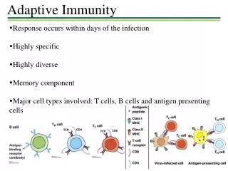
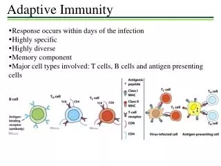
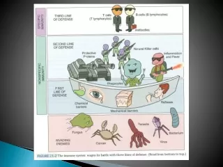
Third Line of Defense • Is called adaptive immunity • The body’s ability to recognize and defend itself against distinct invaders and their products • Is a “smart” system whose “memory” allows it to respond rapidly to a second encounter with a pathogen • Antigens trigger specific immune responses • Various cells, tissues, and organs are part of specific immunity • We will concentrate on B and T lymphocytes
Antigens Quick definition=Molecules that trigger a specific immune response • Include components of bacterial cell walls, capsules, pili, and flagella, as well as proteins of viruses, fungi, and protozoa • Food and dust can also contain antigenic particles • Enter the body by various methods
The Lymphatic System • Screens the tissues of the body for foreign antigens • Composed of lymphatic vessels and lymphatic cells Figure 16.2
Lymphatic Vessels • Form a one-way system that conducts lymph from local tissues and returns it to the circulatory system • Lymph is a liquid with similar composition to blood plasma that arises from fluid leaked from blood vessels into surrounding tissues • An accumulation of too much fluid in tissues is called Edema
Lymphoid Cells • Develop from stem cells in the red bone marrow • Includes lymphocytes, the smallest of the leukocytes
Lymph Nodes • Houses leukocytes that recognize and attack foreign antigens present in the lymph • Concentrated in the cervical (neck), inguinal (groin), axillary (armpit), and abdominal regions • Receives lymph from afferent lymphatic vessels and drains lymph into efferent lymphatic vessels
Other Lymphoid Tissues and Organs • Spleen • Similar in structure and function to the lymph nodes • Filters bacteria, viruses, toxins, and other foreign matter from the blood • Tonsils and mucosa-associated lymphoid tissue (MALT) • Physically trap foreign particles and microbes • MALT includes the appendix, lymphoid tissue of the respiratory tract, vagina, urinary bladder, mammary glands, and Peyer’s patches in the wall of the small intestine
B Lymphocytes • Arise and mature in the red bone marrow • Found primarily in the spleen, lymph nodes, and MALT • Small percentage of B cells circulate in the blood • Major function is the secretion of antibodies
Antibodies • Also called immunoglobulins (Ig) • Soluble proteins that bind antigen • Secreted by plasma cells, which are B cells actively fighting exogenous antigen • Considered part of the humoral immune response since bodily fluids such as lymph and blood were once called humors
Antibody Structure • Antigen-binding sites are complementary to antigenic determinants (epitopes) Figure 16.5
Functions of Antibodies • Function in several ways • Activation of complement • Stimulation of inflammation • Neutralization • Opsonization • Agglutination Figure 16.6
Classes of Antibodies • Apparently a single type of antibody is not sufficient for the multiple types of invaders to the body • The class involved in the immune response depends on the type of foreign antigen, the portal of entry, and the antibody function needed • Five different classes of antibodies
Classes of Antibodies Figure 16.7
B Cell Receptor (BCR) • Is an antibody that remains associated with the cytoplasmic membrane • Each B lymphocyte has multiple copies of a single type of BCR • Antigen binding site is identical to that of the secreted antibody for that particular cell • Each BCR is complementary to only one antigenic determinant • The BCRs on all of an individual person’s B cells are capable of recognizing millions of different antigenic determinants
T Lymphocytes • Produced in the red bone marrow and mature under the influence of the thymus • Circulate in the lymph and blood and migrate to the lymph nodes, spleen, and Peyer’s patches • Part of the cell-mediated immune response because they act directly against various antigens • Cells that harbor intracellular pathogens • Abnormal body cells such as cancer cells that produce abnormal cell surface proteins
T Cell Receptor Figure 16.9
Cytotoxic T cells (TC Cells) • Distinguished by the CD8 cell-surface glycoprotein • Directly kill certain cells • Cells infected with viruses and other intracellular pathogens • Abnormal cells, such as cancer cells
Helper T Cells (TH Cells) • Distinguished by the CD4 cell-surface glycoprotein • Function to “help” regulate the activities of B cells and cytotoxic T cells during an immune response • Secrete various soluble protein messengers, called cytokines, that determine which immune response will be activated
Helper T Cells (TH Cells) • Two types • Type 1 helper T cell (TH1) • Assist cytotoxic T cells • Express a cytokine receptor named CCR5 • Type 2 helper T cell (TH2) • Assist B cells • Have cytokine receptors CCR3 and CCR4
Cytokines • Soluble regulatory proteins that act as intercellular signals when released from certain body cell • Immune system cytokines signal among various leukocytes • The complex web of signals among all the cell types of the immune system is referred to as the cytokine network
Cytokines of the Immune System • Interleukins (ILs) – signal among leukocytes • Interferons (IFNs) – antiviral proteins that may act as cytokines • Growth factors – proteins that stimulate stem cells to divide, maintaining a adequate supply of leukocytes • Tumor necrosis factor (TNF) – Secreted by macrophages and T cells to kill tumor cells and regulate immune responses and inflammation • Chemokines– signal leukocytes to go to a site of inflammation or infection and stimulate other leukocytes
Lymphocyte Editing by Clonal Deletion • Vital that immune responses not be directed against autoantigens • Body “edits” lymphocytes to eliminate any self-reactive cells
Lymphocyte Editing by Clonal Deletion No Match Match Figure 16.10
Major Histocompatibility Complex (MHC) • Important in determining the compatibility of tissues in successful tissue grafting (transplantation) • Major histocompatibility antigens are glycoproteins found in the membranes of most cells of vertebrate animals • Function to hold and position antigenic determinants for presentation to T cells • Antigens bind in the antigen-binding groove of MHC molecules • Two classes of MHC proteins • MHC class I • MHC class II
Antigen Processing • T-independent antigen • Large antigen molecules with readily accessible, repeating antigenic determinants • B cells can bind these directly without being processed • Stimulates B cells to differentiate into a plasma cell and produce antibodies
Antigen Processing • T-dependent antigens • Smaller antigens with less accessible antigenic determinants • B cells require involvement from helper T cells to target these antigens • Helper T cells are assisted by leukocytes that process the antigen to make the antigenic determinants more accessible • Processing is different based on whether the antigen is exogenous or endogenous
Processing of Exogenous Antigens • APC internalizes the invading pathogen and enzymatically digests it into smaller antigenic fragments which are contained within a phagolysosome • Phagolysosome fuses with a vesicle containing MHC II molecules • Each fragment binds to the antigen-binding groove of a complementary MHC II molecule • The fused vesicle then inserts the MHC II-antigen complex into the cytoplasmic membrane so the antigen is presented on the outside of the cell
Processing of Endogenous Antigens • The intracellular pathogens are also digested into smaller antigenic determinants • Each fragment binds to a MHC I molecule located in the endoplasmic reticulum membrane • The membrane is packaged into a vesicle by a Golgi body which is inserted into the cytoplasmic membrane so the antigen is displayed on the cell’s surface Antigen presentation movie (3)
Humoral Immune Response • Body mounts humoral immune responses against exogenous pathogens • Components of a humoral immune response • B cell activation and clonal selection • Memory B cells and the establishment of immunological memory Animation: Humoral Immunity PLAY
Plasma Cells • Make up the majority of cells produced during B cell proliferation • Each plasma cell secretes only antibody molecules complementary to the specific antigenic determinant • Are short-lived cells that die within a few days of activation, though their antibodies and progeny can persist
Memory B Cells • Cells produced by B cell proliferation that do not secrete antibodies • Long-lived cells that divide only a few times and then persist in the lymphoid tissue • Are available to initiate antibody production if the same antigen is encountered again
Primary and Secondary Responses Video (part 3)
Cell-Mediated Immune Response • Responds to intracellular pathogens and abnormal body cells • The most common intracellular pathogens are viruses but the response is also effective against intracellular bacteria • Triggered when antigenic determinants of the pathogen are displayed on the host cell’s surface • Cytotoxic T-cells secrete perforin and granzyme that lead to the destruction of the target cell.
A Cell-Mediated Immune Response Animation: Cell-Mediated Immunity PLAY Figure 16.17
Acquired Immunity • Specific immunity acquired during an individuals life • Two types • Naturally acquired- immune response against antigens encountered in daily life • Artificially acquired- response to antigens introduced via a vaccine • Further distinguished as either active or passive • Active- active response to antigens via humoral or cell-mediated responses • Passive- passively receive antibodies from another individual