Download
1 / 74
750 likes | 1.11k Vues
Explore the characteristics and subtypes of T cells in adaptive immunity, including helper, cytotoxic, memory, and regulatory cells. Learn how memory T cells provide long-term immunity and how regulatory T cells prevent autoimmunity.

E N D
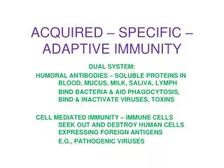
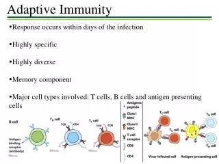
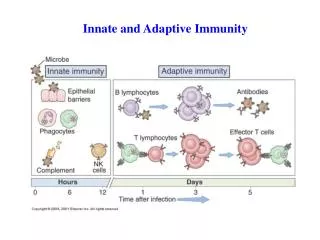
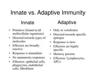
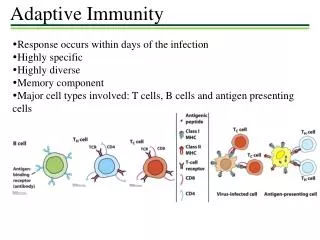
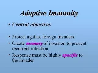
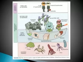
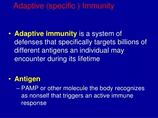
Adaptive (Acquired) Immunity characteristics: • Generation of antigen – specific lymphocytes “effector cells” • Generation of Memory cells which prevent reinfection with the same organism. • It takes more time to develop “>96 h” • It exhibits specificity against foreign substances. • Consists from cellular (lymphocytes) and humeral (antibodies) immunity
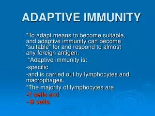
T cells • A T cell, or T lymphocyte, is a type of lymphocyte that plays a central role in cell-mediated immunity. • T cells can be distinguished from other lymphocytes, such as B cells and natural killer cells, by the presence of a T-cell receptor on the cell surface. • They are called T cells because they mature in the thymus from thymocytes (although some also mature in the tonsils). • The several subsets of T cells each have a distinct function.
T cell types • Helper (CD4+) • Cytotoxic or Killer (CD8+) • Memory • Regulatory (suppressor) • Natural killer T cell • Mucosal associated invariant • Gamma delta T cells
T helper cells (TH cells) • They assist other white blood cells in immunologic processes, including maturation of B cells into plasma cells and memory B cells and activation of cytotoxic T cells and macrophages. • These cells are also known as CD4+ T cells because they express the CD4 glycoprotein on their surfaces. • Helper T cells become activated when they are presented with peptide antigens by MHC class II molecules, which are expressed on the surface of antigen-presenting cells (APCs).
T helper cells (TH cells) • Once activated, they divide rapidly and secrete small proteins called cytokines that regulate or assist in the active immune response. • These cells can differentiate into one of several subtypes, including TH1, TH2, TH3, TH17, TH9, or TFH, which secrete different cytokines to facilitate different types of immune responses. • Signaling from the APC directs T cells into particular subtypes
Transcription factors • Intracellular pathogens • Macrophage activation Cytokine secreted by APC • Extracellular pathogens • Antibodies production • Helminths and allergy • Autoimmunity • Tumor suppression • Extracellular bacteria and fungi • Neutrophils activation • promote tissue repair or remodeling. pathogenesis of inflammatory skin disorder • Long term humoral immunity • Inhibit dendritic cells • Suppress T cells • Prevent autoimmunity
T cytotoxic • Cytotoxic T cells (TC cells, CTLs, T-killer cells, killer T cells) destroy virus-infected cells and tumor cells, and are also implicated in transplant rejection. • These cells are also known as CD8+ T cells since they express the CD8 glycoprotein at their surfaces. • These cells recognize their targets by binding to antigen associated with MHC class I molecules, which are present on the surface of all nucleated cells. • Through IL-10, adenosine, and other molecules secreted by regulatory T cells, the CD8+ cells can be inactivated to an anergic state, which prevents autoimmune diseases.
Memory T cells • The single unifying theme for all memory T cell subtypes is that they are long-lived and can quickly expand to large numbers of effector T cells upon re-exposure to their cognate antigen. By this mechanism they provide the immune system with "memory" against previously encountered pathogens. Memory T cells may be either CD4+ or CD8+ and usually express CD45RO. • Memory T cell common subtypes: • Central memory T cells (TCM cells) express CD45RO, C-C chemokine receptor type 7 (CCR7), and L-selectin (CD62L). Central memory T cells also have intermediate to high expression of CD44. This memory subpopulation is commonly found in the lymph nodes and in the peripheral circulation.
Effector memory T cells (TEM cells) express CD45RO but lack expression of CCR7 and L-selectin. They also have intermediate to high expression of CD44. These memory T cells lack lymph node-homing receptors and are thus found in the peripheral circulation and tissues. • Tissue resident memory T cells (TRM) occupy tissues (skin, lung, gastrointestinal tract, etc.) without recirculating. One cell surface marker that has been associated with TRM is the integrin αeβ7. These cells are thought to play a major role in protective immunity against pathogens. Dysfunctional TRM cells have been implicated in autoimmune diseases, such as psoriasis, rheumatoid arthritis, inflammatory bowel disease.
Virtual memory T cells (TVM) differ from the other memory subsets in that they do not originate following a strong clonal expansion event. Thus, although this population as a whole is abundant within the peripheral circulation, individual virtual memory T cell clones reside at relatively low frequencies.
Regulatory T cells (suppressor T cells) • Their major role is to shut down T cell-mediated immunity toward the end of an immune reaction and to suppress autoreactive T cells that escaped the process of negative selection in the thymus. • Crucial for the maintenance of immunological tolerance. • Regulatory T cells can develop either during normal development in the thymus, and are then known as thymic Treg cells, or can be induced peripherally and are called peripherally derived Treg cells. These two subsets were previously called "naturally occurring", and "adaptive" or "induced", respectively.
Both subsets require the expression of the transcription factor FOXP3 which can be used to identify the cells. Mutations of the FOXP3 gene can prevent regulatory T cell development, causing the fatal autoimmune disease (IPEX). • Several other types of T cell have suppressive activity, but do not express FOXP3. These include Tr1 cells and Th3 cells, which are thought to originate during an immune response and act by producing suppressive molecules. Tr1 cells are associated with IL-10, and Th3 cells are associated with TGF-beta. Recently, Treg17 cells have been added to this list.
Natural killer T cell • Natural killer T cells bridge the adaptive immune system with the innate immune system. • Unlike conventional T cells that recognize peptide antigens presented by major histocompatibility complex (MHC) molecules, NKT cells recognize glycolipid antigen presented by a molecule called CD1d.
Natural killer T cell • Once activated, these cells can perform functions ascribed to both Th and Tc cells (i.e., cytokine production and release of cytolytic/cell killing molecules). • They are also able to recognize and eliminate some tumor cells and cells infected with herpes viruses. • (NKT cells – not to be confused with natural killer cells of the innate immune system which is CD3 negative).
Gamma delta T cells • The majority of human T cells, termed alpha beta T cells (αβ T cells), rearrange their alpha and beta chains on the cell receptor and are part of the adaptive immune system. Specialized gamma delta T cells, (a small minority of T cells in the human body, more frequent in ruminants), have invariant T-cell receptors with limited diversity, that can effectively present antigens to other T cells and are considered to be part of the innate immune system. • This group of T cells is much less common in humans and mice (about 2% of total T cells)
B cells • also known as B lymphocytes, are a type of white blood cell of the lymphocyte subtype. • They function in the humoral immunity component of the adaptive immune system by secreting antibodies. • Additionally, B cells present antigen (they are also classified as professional antigen-presenting cells (APCs)) and secrete cytokines. • In mammals, B cells mature in the bone marrow, which is at the core of most bones
B cells • In birds, B cells mature in the bursa of Fabricius, a lymphoid organ. (The "B" from B cells comes from the name of this organ, where it was first discovered by Chang and Glick, and not from bone marrow as commonly believed). • B cells, unlike the other two classes of lymphocytes, T cells and natural killer cells, express B cell receptors (BCRs) on their cell membrane. BCRs allow the B cell to bind to a specific antigen, against which it will initiate an antibody response. • B cells have many subclasses. Brief info about them could be found in the slide notes.
IL-21 & others
What is an antibody? • Product of adaptive immunity • Made specifically to bind a unique antigenic epitope (also called an antigenic determinant) • Possesses an antigen binding site • A product of the Plasma cell • Members of the class of proteins called immunoglobulins
Fab VH VL CH1 CL CH2 Fc CH3 Immunoglobulin G (IgG) • 2 Heavy and 2 Light chains • 2 Fab and 1 Fc fragment • 4 Subclasses (IgG1, IgG2, IgG3, IgG4) • Mol. Wt. 150,000 • ~70-75% of serum immunoglobulin. • The major antibody of the secondary immune response • Change in affinity with time
J-Chain Immunoglobulin M (IgM) • 10 Heavy and 10 Light chains • 10 Fab and 5 Fc fragments • Mol. Wt. ~900,000 • <10% of serum immunoglobulin. • Single J Chain (15 kDA) • The predominant "early" antibody • Most primitive immunoglobulin • No change in affinity with time
Immunoglobulin A (IgA) • Monomeric- Serum IgA • ~15-20% of serum immunoglobulins • 2 Heavy and 2 Light • 2 Subclasses (IgA1 and IgA2) • Found in serum
Secretory Immunoglobulin A (sIgA) • 4 heavy chains and 4 light chains (dimeric) • J-Chain and secretory component • The major immunoglobulin of secretions • Not found in serum J-Chain Secretory Component
Immunoglobulin E (IgE) • 2 Heavy and 2 Light chains • Mol. Wt. ~190,000 • Trace serum protein • Note CH4 region on H chain • Associated with atopic or anaphylactic hypersensitivity • May play role in immunity to helminthic parasites CH4
Immunoglobulin D (IgD) • 2 heavy and 2 light chains • <1% of serum immunoglobulins • Serves as a membrane receptor on B lymphocytes • May play role in antigen-stimulated lymphocyte differentiation
LYMPHATIC ORGANS • These organs are the sites in which lymphocyte maturation, differentiation and proliferation take place. • The primary “central” lymphoid organs... Thymus gland and bone marrow. Maturation of T and B lymphocytes into antigen – recognizing lymphocytes and acquiring their antigen. • Secondary “peripheral” lymphoid organs • They are those organs where antigen–driven proliferation and differentiation occur. These include lymph nodes, spleen, gut-associated lymphoid tissues such as tonsils.
Thymus Gland • Progenitor cells from the bone marrow migrate to the thymus, where they differentiate into T lymphocytes • The thymus gland is a lymphoepitheloid bilobed structure • It is derived from the endoderm of the third and fourth pharyngeal pouches. • During fetal development, the size of the thymus increases, the growth continues until puberty, thereafter, the thymus undergoes atrophy with aging. • The cortex and medulla are infiltrated with thymocytes. T lymphocytes mature in cortex and migrate to the medulla. Macrophages and dendritic cells are found in medulla.
Macrophages are involved in clearing apoptotic thymocytes. • Mature T lymphocytes migrate to the secondary lymphoid organs in which they encounter and respond to foreign antigens. • T lymphocytes recognize and respond to foreign antigen via their specific receptors “TCR” which is acquired during differentiation in the thymus. • Only 5-10% of maturing lymphocytes survive and eventually leave the thymus, 90-95% of all thymocytes die in the thymus. • Lymphocytes which die have developed specificity to self structures or have failed to make functional receptors and, therefore, are eliminated.
Bone marrow • In embryonic life , B cells differentiate from hematopoietic stem cells in the fetal liver. • After birth this function moves to the bone marrow. • B cells mature in bone marrow and bear antigen – specific receptors that have a structure and specificity identical to the antibody later synthesize by that B cells. • The mature B cells are transported by the circulating blood to the secondary lymphoid organs, where they encounter and respond to foreign antigens.
Secondary Lymphoid Organs • The major secondary lymphoid organs are the spleen and lymph nodes. In addition, tonsils, appendix, peyer’s patches, and lymphoid aggregates spread throughout mucosal tissue “mucosal – associated lymphoid tissue – MALT”. Those associated with the gut “gut – associated lymphoid tissue – GALT, bronchus – associated lymphoid tissue BALT” • The secondary lymphoid organs have two major functions: • Trapping and concentrating foreign substances • The main sites of production of antibodies and the induction of antigen – specific T Lymphocytes.
The spleen • The largest organ of the secondary lymphoid organs. • Traps and concentrates foreign substances carried in the blood. • The major organ of antibody synthesis • White pulp is rich in lymphoid cells • Red pulp contains many sinuses, large quantities of RBCs and macrophages , some lymphocytes.
The spleen • B – cells are present mainly in germinal centers, T cells in the peripheral region. • Approximately 50% of spleen cells are B lymphocytes, 30-40% are T lymphocytes. • After antigenic stimulation, the germinal centers contain large numbers of B cells and plasma cells, these cells synthesize and release antibodies.
Lymph nodes • lymph nodes are small, ovoid structures • They are close to major junction of the lymphatic channels, which are connected to the thoracic duct. • The thoracic duct transports lymph and lymphocytes to the vena cava, the vessel that carries blood to the right side of the heart, from where it is redistributed throughout the body. • The cortical region contains primary lymphoid follicles.
Lymph nodes • After antigenic stimulation, they enlarge to form secondary lymphoid follicles with germinal centers containing B lymphocytes. They generate clones of cells with higher affinity receptors “antibody” for the antigenic epitope that triggered the initial response. • The deep cortical area or Para cortical region contains T cells and dendritic cells which present antigen fragments to T cells • The medullary area of the lymph node contains antibody – secreting plasma cells that have travelled from the cortex to the medulla via lymphatic vessels.
The migration of lymphocytes between various lymphoid and nonlymphoid tissue and their homing to a particular site is highly regulated by means of various cell – surface adhesion molecules “CAMs” and receptors to these molecules. • Blood lymphocytes cross the endothelial vascular lining of post capillary vascular sites termed high endothelial venules “HEVs” this process is called extravasation. • Recirculating lymphocytes selectively bind to specific receptors on the HEV of lymphoid tissue. • Recirculating monocytes and granulocytes also express adhesion molecule receptors and migrate to tissue sites using a similar mechanism.