Download
1 / 64
670 likes | 1.22k Vues
Viral Haemorrhagic Fevers. Craig Corcoran NHLS Virology, Groote Schuur Hospital. VHF- what is it all about?. VHF’s attract the attention of medical professionals and the general public for a variety of reasons

E N D
Viral Haemorrhagic Fevers Craig Corcoran NHLS Virology, Groote Schuur Hospital
VHF- what is it all about? • VHF’s attract the attention of medical professionals and the general public for a variety of reasons • They are high on the public mind as they are thought of as highly infectious, killing their victims in a dramatic way • Mysteries remain as to the source of some of them
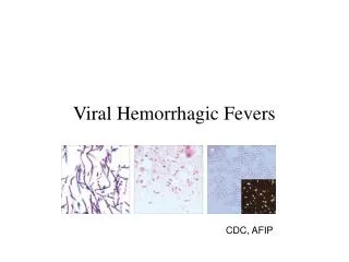
“Viral Haemorrhagic Fever” An acute febrile illness characterized by malaise, myalgia, and prostration dominated by generalized abnormalities of vascular permeability, and regulation. Bleeding manifestations often occur, particularly in severe cases; they are usually diffuse and reflect widespread vascular damage rather than life-threatening volume loss.
Flaviviridae (dengue, yellow fever, TBE encephalitides) Viral Haemorrhagic Fevers Arenaviridae (Lassa, Junin, Machupo, Guanarito) Enveloped RNA viruses Bunyaviridae (CCHF, RVF, Hantaviruses) Filoviridae (Ebola, Marburg)
These viruses share a number of features: • They are all RNA viruses and are enveloped (i.e covered in a fatty (lipid) coating • Their survival is dependent on an animal or insect host called the natural reservoir • They are geographically restricted to areas where their host species live • Humans are not the natural reservoir for any of these viruses. Humans are infected when they come into contact with infected hosts, and with some viruses, can transmit the virus to one another • Human outbreaks occur sporadically and irregularly. These outbreaks cannot be easily predicted • With few exceptions, there is no cure or established drug treatment for VHFs
Early clinical signs and symptoms may be very discrete and cannot easily be distinguished from those of other illnesses
Clinical signs and symptoms are easier to interpret once the disease has progressed already
VHF-clinical picture • Short incubation period • Non-specific onset of illness • Headache, myalgia, arthralgia • Pharyngitis, conjunctival injection/bleed • GIT discomfort/disturbances • Impaired consciousness • Haemorrhages • Proteinuria • Jaundice • Rash, exanthema
VHF-differential diagnosis • VHF vs. VHF: • clinical picture-unreliable, epidemiology-approximate, laboratory-proof • VHF vs. bacterial infections • Typhoid, leptospirosis, tick-bite fever, shigellosis, purulent pharyngitis, sepsis (streptococcal, staphylococcal, meningococcal), plague • VHF vs. parasitic diseases • Malaria, african trypanosomiasis, amoebiasis • VHF vs. viral diseases • Viral hepatitis, herpes simplex
Dengue fever • Main hosts- non human primates • Human-to-human transmission through Aedes spp. • 2.5 billion individuals at risk • 40-80 million infected each year with thousands of deaths
Dengue-clinical features • Fever, headache, back pain , chills, musculoskeletal pain, rash, leucopaenia, thrombocytopaenia • Usually lasts 4-10 days • Dengue haemorrhagic fever/Dengue shock syndrome • Acute vascular hyperpermeability plus abnormal haemostasis • Rapid deterioration after 2-5 days • Scattered petechiae, ecchymoses, easy bruising/bleeding, hepatomegaly, epigastric pain • Pathogenesis: enhancing antibodies- maternal in infants, second infection with a different serotype • Supportive treatment, vaccine in development
Yellow Fever • Historic illness stretching back 400 years • yellow: jaundice affecting certain patients • Mosquitos (Aedes and haemogogus) are the true reservoir and vector • Estimated 200 000 cases/year, 30 000 deaths • Symptoms vary from mild to severe with haemorrhagic manifestations Africa and South America only
‘acute’ phase- fever, headache, muscle pain, GIT disturbance • 15% enter a ‘toxic’ phase and rapidly develop jaundice with bleeding manifestations and renal failure. 50% die within 10-14 days • Supportive treatment • Prevention: vaccine- 17D live attenuated, safe and highly effective
Filoviruses: Ebola HF • 1976- Simultaneous large outbreaks in Yambuku (Zaire, now DRC) and Nzara/Maridi (Sudan) • Originally thought to be one outbreak • Virology now recognises 2 distinct viruses • EBO-Z: 318 cases; 88% fatal • EBO-S: 284 cases; 53% fatal
Ebola Outbreaks 1979, 2004 1994 1976, 1979, 2004 1994, 1996, 1996 2000 Congo 2003 1976, 1995 *Doctor returning from Gabon 1996*
Filoviruses: Marburg HF • 1967: Marburg, Frankfurt & Belgrade • African green monkeys from Uganda • 25 primary • 6 secondary • 1 sexual transmission from husband to wife 85 days after onset of illness, virus cultured from semen • 7 deaths
Routes of transmission: filoviruses • Contact with body fluids of an ill patient • HCW and relatives • Infected carcasses (handling/cutting of dead primates) • Needle transfer • Preparation of body for burial • Sexual transmission • Laboratory accident • Aerosol infectivity potential demonstrated experimentally in monkeys (Ebola)
Reservoir of infection • Not identified in terrestrial animals or in insects • Non-human primates suffer but are not the reservoir • Association with caves and mines make bats suspects for Marburg • Fruit bats- ? reservoir for Ebola and Marburg (antibodies and RNA found by researchers in Gabon)
Filoviruses: clinical presentation • 1-2 week incubation • Abrupt onset fever, headache, myalgia • Non-pruritic papular erythematous eruption becoming large coalescing macules and papules • Palatal petechiae and haemorrhages • GI symptoms, chest pain, delirium • Sever cases- haemorrhages from venipuncture sites, mucous membranes and venipuncture sites • 53-88% case-fatality • ~ 45% hemorrhage • Supportive treatment • Vaccines in development
Marburg blanching maculopapular rash, day 5, Johannesburg 1975
Arenaviridae • Arenaviruses associated with human disease VirusOrigin of NameYearDistribution Lassa Town, Nigeria 1969 West Africa Junin Town, Argentina1957 South America Machupo River, Bolivia 1962 South America Guanarito Area, Venezuela 1989 South America Sabia Town, Brazil 1990 South America LCMV Clinical disease 1933 Worldwide
Lassa: general facts • Viral hemorrhagic fever caused by the Arenavirus Lassa • Transmitted from rodents to humans • Discovered in Nigeria, 1969 • Endemic in portions of West Africa • Seasonal clustering: Late rainy and early dry season • Affects all age groups and both sexes
Lassa virus “arenosus” (Latin “sandy”)
Endemic in areas of West Africa, including Nigeria, Liberia, Sierra Leone, and Guinea • Estimated 300,000-500,000 infections/year, with 5000 deaths • Rodent-to-human transmission (the “multimammate rat”, Mastomys species-complex) • Secondary human-to-human transmission with the potential for nosocomial outbreaks with high case-fatality
Rodent reservoir Mastomysspecies complex
Lassa: Transmission • Rodent-to-human: • Inhalation of aerosolized virus • Ingestion of food or materials contaminated by infected rodent excreta • Catching and preparing Mastomys as a food source
Lassa: Transmission • Human-to-human: • Direct contact with blood, tissues, secretions or excretions of infected humans • Needlestick or cut • Inhalation of aerosolized virus • Sex • Breast feeding
Lassa: Clinical Aspects • 80% asymptomatic • Incubation period of 5-21 days • Gradual onset of fever, headache, malaise and other non-specific signs and symptoms • Pharyngitis, myalgias, retro-sternal pain, cough and gastrointestinal symptoms typically seen • A minority present with classic symptoms of bleeding, neck/facial swelling and shock • Case fatality of hospitalized cases: 15-20% • Particularly severe in pregnant women and their offspring • Deafness a common sequela
Lassa: Treatment • Supportive measures • Ribavirin • Guanosine nucleoside analog: blocks viral replication by inhibiting IMP dehydrogenase • Licensed for treatment of RSV and HCV • Potential adverse effects: • Dose dependent reversible anemia • Pancreatitis • Teratogen in rodents
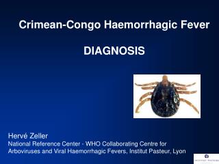
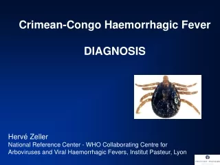
CCHF-some background • 1944- Crimean peninsula- Crimean haemorrhagic fever (about 200 cases) • 1956- Belgian Congo- 1 child- Congo Fever • Virus isolated in suckling mice in 1967 • 1-10 cases diagnosed annually in South Africa • Case fatality rate 20-25%, 30-50% without proper medical attention • Mid 1980’s- nosocomial outbreak at TBH- 8 cases, 2 deaths • 27 cases October 1996- Oudtshoorn ostrich abattoir workers
Distribution of the bont-legged ticks in South Africa • reservoir and vector Hyalomma marginatum rufipes Hyalomma marginatum turanicum Hyalomma truncatum
Hyalommas are two host ticks • Lavae and nymphs feed on the first host • Adults feed on the second host • Cattle • Sheep • Goats • Ostriches
So when are humans at risk? • Bitten by tick/s or crushed tick/s with bare hands • Direct contact with fresh blood or other tissues of livestock or game animals (ear tagging, castration ect.) • Direct contact with blood, secretions or excretions of a confirmed or suspected CCHF patient including needlestick injuries • Resided in or visited a rural environment where contact with livestock or ticks was possible but a specific incident constituting exposure cannot be identified • NB- incubation period usually 2-7 days hence exposure usually < 7days
What are the clinical features? • Sudden onset • Fever ≥ 38ºC on at least one occasion • Severe headache • Myalgia • Nausea and/or vomiting • Pharyngitis, conjunctivitis • Bleeding tendency: petechial rash, ecchymoses, epistaxis, haematemesis, haematuria or melaena
CCHF- laboratory findings • Leukopaenia or leukocytosis • WCC< 3 x 10⁹/l or ≥ 9 x 10⁹/l • Thrombocytopaenia • Platelet < 150 x 109/l • Usually < 100 x 109/l • Abnormal INR and APTT • Transaminitis • AST ≥ 100iu/l • ALT ≥ 100iu/l
CCHF-differential diagnosis • Malaria, tick bite fever, disseminated HSV, viral hepatitis, typhoid, rift valley fever, anthrax, brucellosis, Q fever… • History of exposure, incubation period following exposure, signs and symptoms, laboratory findings
CCHF : viral/antibody kinetics IgM IgG viremia 0 5 10 RT-PCR 16 Viral isolation ELISA IgM IgG IFA IgM duration: 2-3 months up to 6 months…