Hemorrhagic Fevers
820 likes | 1.6k Vues
Hemorrhagic Fevers With a Concentration on Filoviruses Simon A Francis Susanna Epstein Medha Goyal History Hemorrhagic Fevers Definition: a severe multi-system syndrome . Vascular System Damaged Body regulation Impaired Accompanied by hemorrhage Four families of viruses

Hemorrhagic Fevers
E N D
Presentation Transcript
Hemorrhagic Fevers With a Concentration on Filoviruses Simon A Francis Susanna Epstein Medha Goyal
History • Hemorrhagic Fevers • Definition: a severe multi-system syndrome. • Vascular System Damaged • Body regulation Impaired • Accompanied by hemorrhage • Four families of viruses • Arenaviruses (Junin Virus) • Bunyaviruses (Nariovirus ) • Flaviviruses (ie. Yellow Fever) • Filoviruses (Marburg & Ebola) • CDC Classification: BSL-4 Agent
History • Hemorrhagic Fevers (Continued) • RNA viruses covered in a lipid coating • Viruses are geographically restricted to areas where host species live • Humans are not natural reservoirs for these viruses
History • Filoviruses • Marburg Virus • A.K.A. (African Hemorrhagic Fever, Green Monkey Disease, Marburg Fever) • First outbreak (Marburg Germany, 1967) • laboratory workers infected by monkeys • Simultaneous outbreak in Hamburg, Germany and Belgrade, Yugoslavia (now Serbia) • 32 human cases • 31 primary one generation of secondary transmission • 23% of human mortality • Overall Mortality 23%-25%
History • Filoviruses (continued) • Ebola Virus • Named after a river in the Republic of Congo (Formerly Zaire) • First outbreak (Zaire 1967) • 318 human cases • 88% mortality • Disease spread by close personal contact in hospital setting (amplification) • Fatal in humans and non-human primates • Four subtypes (~80 nm in diameter) • Ebola-Zaire(990-1086 nm in length ), Ebola-Sudan (974-1063 nm in length), Ebola-Ivory Coast, Ebola-Reston (disease in non-human primates 1026-1083 nm in length )
History • Filoviruses (Continued) • Ebola (Continued) • Sudan international scientist arrived • To late to deal with virulent epidemics • Hospitals closed • Infected patients quarantined • Reconstructed data from survivors
Outbreaks • Marburg • Europe Outbreaks • 1967 • Hamburg and Marburg, Germany and Belgrade, Yugoslavia • Africa Outbreaks • 1975 • Johannesburg, South Africa • 3 died • 1980 • Western Kenya • 2 Died (Physician died in Nairobi) • 1987 • Young man traveling extensively in Kenya • 1999-2000 • Outbreak in Durba, Republic of Congo • Cases linked to workers in a gold mine
Outbreaks • Ebola (types named after location) This diagram was adapted from a WHO publication accompanied by the following note (edited): Phylogenic tree showing the evolutionary relationship of Ebola viruses (courtesy of A. Sanchez, Centers for Disease Control and Prevention [CDC]; derived from Georges-Courbet MC, Sanchez A, Lu CY, et al. Isolation and phylogenetic characterization of Ebola viruses causing different outbreaks in Gabon. Emerg Infect Dis 1997;3:59-62).
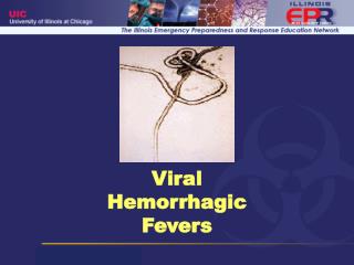
New York Times • February 15, 2003 • World Briefing: Africa • CONGO REPUBLIC: EBOLA TOLL REACHES 51 The death toll from a suspected outbreak of the deadly Ebola virus in Congo Republic has crept up to 51, and people have begun fleeing into dense forest to escape what some believe to be an evil spell. The authoritieshave tried to impose tight restrictions on movement in the hope of preventing the spread of the outbreak, the second reported in little over a year in the country's remote northwest. It is thought to have been caused by the consumption of infected monkey meat. (Reuters)
Transmission • Ebola Virus • No carrier state (reservoir Unknown) • Researchers Hypothesize that it is Zoonotic • Unpopular theory that plant may be the reservoir of the virus • Human to Human Transmission through contact of contaminated secretions. • Ebola-Reston • Occurred in the U.S (Reston, VA) • Occurred in African Green Monkeys • Why only to non-human primates? • Four scientist found to have antibody for the disease • Circumstantial Evidence of airborne transmission • Spread within and between rooms (national center for Infectious Diseases) • Marburg Virus • Transmission from animal host unknown • Human to Human (Close Contact and change of fluids highly suspect source of transmission)
Reservoir • Not known • May be • A rare species • One that usually does not contact clinical host • If contact is made the virus may not be easily transmitted • Hypothesize • Bats • Plants
Filoviruses General Facts • Replication: • Not fully understood. Created by budding of the surface of their host cells (Susanna) • Structure: • Pleomorphic: Long sometimes branched filament shaped like a “6”, “U” or a circle. • Each Virion contains one molecule of single stranded, negative sense RNA (Susanna) • Symptoms of Ebola and Marburg (Medha)
INCUBATION & DEATH PERIODS • Ebola Incubation at 2 – 21 Days • Marburg Incubation: : 3-9 days • VHF (in general) - Death Between 7 – 16 Days • Ebola Death Ensues as Early as 2 Days after expression of symptoms
Morphology and Structure • Filamentous or Bacillus form • Vary greatly in length (up to 14000 nm) Mean Unit Length Marburg- 860 nm Ebola- 1200 nm • Uniform diameter of 80 nm
Filovirus Composed of: • Ribonucleoprotein complex (nucleocapsid) • Matrix • Envelope studded with peplomers (10 nm long)
Filovirus Genome • Nonsegmented, negative-strand RNA • Filovirus Genome – 19 kb • 7 Genes • Nucleotide and amino acid differences b/w Marburg and Ebola – 55% Ebola – 37 to 41% • Overlaps 1 in Marburg genome – VP30 to VP24 2 in Ebola genome- VP35 to VP40 & GP to VP30
Glycoprotein Gene Marburg- GP gene encodes 1 product 1 open reading frame (0) Ebola- GP gene encodes 2 products 2 open reading frames (0 & -1) connected by the insertion of 1 additional A at a series of 7 U’s on the genomic RNA
The 7 sequentially arranged genes are transcribed into Ebola - 8 major mRNAs (7 structural proteins and 1 nonstructural protein) Marburg – 7 major mRNAs (7 structural proteins)
Filovirus Proteins • Ebola and Marburg encode 7 structural proteins • Ebola encodes 1 nonstructural protein • Two Main Categories Associated with the nucleocapsid transcription and replication of viral genome Associated with the envelope assembly of virus receptor binding and virus entry
Ribonucleocapsid Proteins 1. Nucleoprotein (NP) Gene 1 2. Viral Protein 35 (VP35) Gene 2 3. Viral Protein 30 (VP30) Gene 5 4. Polymerase L Gene 7 • Matrix Proteins 5. Viral Protein 40 (VP40) Gene 3 6. Viral Protein 24 (VP24) Gene 6 • Membrane Protein 7. Glycoproteins (GP) Gene 4 • Secreted Protein 8. Secretory Glycoprotein (sGP) Gene 4
Nucleoprotein • Primary structural protein associated with the nucleocapsid • Hydrophobic N-terminal half Binds genomic RNA • Hydrophilic C-terminal half (Variable b/w Marburg and Ebola) Interacts with matrix proteins
VP30- Minor structural protein associated with the nucleocapsid • Polymerase L- Transcription and Replication (largest and least abundant protein) • VP35- Cofactor in transcription and Replication (Cofactor in polymerase complex) • VP40- Matrix protein Virus assembly and budding Forms hexamers when it contacts the plasma membrane which confers added stability during assemebly. (most abundant protein) • VP24- Minor Matrix Protein Possibly uncoats virus during infection
Expression of Glycoprotein • Transcriptional RNA Editing (occurs only in Ebola) At a series of 7 U’s on the genomic RNA template insert a non-template-coded adenosine 20% of GP mRNA is edited GP with 680 amino acids 80% of GP mRNA is not edited sGP with 370 amino acids
GP (Structural) • Formation of GP1-GP2 Heterodimer In the trans-Golgi, the precursor molecule (GP0) is post-translationally cleaved by furin at yielding a heterodimer, (GP1-GP2) Furin cleavage site (Arg-Arg-X-Arg/LYS-Arg) (Marburg, cleavage site is more toward N-terminus) Heterodimer is linked together by one disulfide bond, a cysteine bridge
GP1 Molecule C-terminus: hydrophilic, highly glycosylated Sequences for receptor recognition and binding N-terminus: hydrophobic Connects GP1 to GP2 by a disulfide bond • GP2 Molecule Fusion peptide near near its N-terminus Capable of inserting itself in plasma membranes Believed to mediate the fusion of the host and virus membranes • Functions of GP Forms the Virion Peplomers (Surface Spikes) Trimers of the GP1-GP2 Heterodimer, probably assembled in the ER Mediates viral entry by receptor binding and membrane fusion
sGP (non-structural) • Formation and Structure Homodimer Synthesized from GP mRNA using the conventional ORF Produced from a precursor molecule cleaved by furin near the C-terminus, Precursor molecule SGP and Delta Peptide Homodimer is linked in anti-parallel orientation by 2 disulfide bonds between the 1st and 6th cysteines on separate molecules
Virus Entry and Replicationin Host Cells • Viral surface spikes recognize and bindsurface receptors of host • Virus enters cell via endocytosis • Release of nucleocapsid into cytoplasm • Transcription viral RNA polyadenylated, monocistronic mRNA • Translation and buildup of viral proteins, primarily NP • Budding and release of viruses • Host Cell – dies intracytoplasmic vesiculation, mitochondrial swelling, organelle breakdown
Molecules mediating filovirus entry Marburg • Asialoglycoprotein Receptor (Found exclusively in hepatocytes) Recognizes glycoproteins displaying N-linked sugar chains with terminal galactose residues Ebola • Integrins N-glycosylated transmembrane cell surface receptors Ebola and Marburg • Human folate receptor- Co-factor expressed on cell-surface
Reverse TranscriptionSystem Volchkov et al. contructed 2 recombinant EBOV clones 1. pFL-EBOVe+ antigenomic cDNA clone with authentic editing site 2. pFL-EBOVe- antigenomic cDNA clone with mutated editing site Eliminated editing site using site-directed mutagenesis Editing site in middle of GP gene AAAAAAA EBOV polyadenylation signal ATTAAGAAAAAA AAAAAAA AAGAAGAA
Observations 1. Both pFL-EBOVe+and pFL-EBOVe- Showed typical filovirus structure Possessed similar infectivity and virus production 2. Visible Cytopathic Effects pFL-EBOVe+: 4-6 days after infection But there was still an intact monolayer at day 8 pFL-EBOVe- : 3-4 days after infection 5-6 days after infection, cell rounding was complete
3. Differences in expression of the GP gene Wild Type – 1/5 GP, 4/5 sGP pFL-EBOVe+- 1/5 GP, 4/5 sGP pFL-EBOVe- - no sGP expression, increase in GP expression The increase in GP expression by pFL-EBOVe- no simultaneous increase in virus release most of GP synthesized were immature precursors -with sugar side chains high in mannose -sensitive to treatment with endoglycosidase H -GP transport was arrested in ER or early Golgi
Conclusions • No Transcriptional RNA Editing Over-expression of GP Exhausts Cell Host Machinery Eventual Cell Death • So GP expression and cytotoxicity can be down-regulated by virus through transcriptional RNA editing and sGP expression
Pathogenesis of EBOV Infection • sGP 1. Inhibits early activation of neutrophils -Binds to neutrophils via CD16b cell surface receptor -CD16b activates neutrophils via lateral membrane interaction with CR3 2. Adsorbs neutralizing antibodies • GP 1. Specific region of GP induces cytotoxic effects in endothelial cells -Rapid release of vasoactive agents from infected cells -Induces cell rounding and detachment from extracellular matices -Increases cell membrane premeability
2. Proteolytic activation of GP0 precursor via cleavage -EBO-Z GP cleaved by furin -Prerequisite for fusion between viral envelope and host cell membrane -Enables virus to replicate in host systematic infection 3. Two sequences contribute to evasion of host immunity -Possible immunosuppressive sequence in GP2 molecule -Amino acid sequence at amino terminus suppresses lymphocyte mitogen-stimulated proliferation in vitro
Destruction of the Immune System 1. Infects mononuclear phagocytes and fibroblastic reticular system (associated with lymph nodes) - Failure of early T-cell activation -Disrupts antigen trafficking and cytokine production -Extensive apoptosis of blood leukocytes -Lymphopenia (reduction in lymphocyte #) and severe damage to lymphoid tissue 2. Macrophages and circulating monocytes help transmit virus to other tissues
3. VP35 protein – Type 1 IFN Antagonist -Combats the host interferon response possibly enhancing the replicative ability of the virus