Download
1 / 1
10 likes | 147 Vues
ANAT3231 Cell Biology Neuronal Degeneration in Parkinson’s Disease. Suy, Sophy ;. Introduction. Mitochondrial Dysfunction. [alpha] - Synuclein. Current and future treatments.

E N D
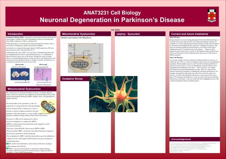
ANAT3231 Cell BiologyNeuronal Degeneration in Parkinson’s Disease Suy, Sophy; Introduction Mitochondrial Dysfunction [alpha] - Synuclein Current and future treatments Parkinson’s Disease (PD) is a progressive neurological disorder involving the basal ganglia. A marked loss in the dopaminergic neurons of the Substantia Nigra (SN) is indicative of PD at a cellular level. Symptomatically it is characterized by important diagnostic features such as rest tremors, bradykinesia, rigidity and postural instability. In Australia, it is estimated that approximately 30,000 people have PD with the greatest prevalence in people aged 50-75. Unfortunately the cause of PD is not clear, but it is thought that genetic and environmental factors (such as environmental toxins), or a combination of them both lead to most cases of the disease. Despite the cause not being known, several pathological processes that may lead to the degeneration of the neurons have been identified. However, definitive research on which process is actually involved has not yet been successful. Macroscopic Microscopic pale SN in PD patients normal pigmented SN normally pigmented SN decreased neurons and pigments in PD patients Drug Therapy:- Drug treatments focus on restoring dopamine levels in the Substantia Nigra or assisting its passage within the brain. Levodopa and dopamine agonists are examples of a current drug therapy. Clinical trials have found some treatments are beneficial in prolonging the life expectancy of Parkinsons patients, with many not showing significant clinical features of the disease if the drug is administered prior to a particular stage in the disease. [2] However, drug therapy has draw backs such as toxicity to the Substantia Nigra, physical side effects and biochemical factors influencing the effectiveness of the drugs in different patients. Stem Cell Therapy:- An exciting example of future treatment of Parkinsons Disease is the use of embryonic stem cells as a source of dopamine neurons that can be transplanted in patients. Mesencephalic precursor cells from the developing midbrain of fetuses have been used as a source of dopaminergic neurons and have been transplanted into Parkinsons rat models. [3] These cells have successfully developed into functional dopaminergic neurons. However, many ethical, medical and practical problems are limiting this treatments potential use. For example, one patient would require the cells from at least five fetuses for success. As well as this dopamine neurons have a poor survival rate of less than 10% and tumour development is a risk associated with stem cell therapy. Despite these drawbacks, some successful trials have highlighted an area with enormous potential in future treatment options. Schematic representation of the MD pathway: . Oxidative Stress Mitochondrial Dysfunction The mechanisms of cell death in Parkinson's disease are largely unknown. Some of the factors that have been implicated in neuronal degeneration in PD include mitochondrial dysfunction (MD), oxidative stress, and aggregation of [alpha]-synuclein. The mitochondira is the 'powerhouse' of the cell responsible for energy production. The mitochondrial electron transport chain is composed of a series of proteins or enzyme complexes attached to the inner membrane of the mitochondria i.e.cristae. In MD, complex-I activity is impaired, resulting in energy failure and the death of DA neurons. • The process of MD can be summarized as follows: • involves the inhibition of complex-I by MPTP • MPTP is a potent neurotoxin captured into acidic organelles, mostly lysosomes of astrocytes. • Astrocytes contain MAO-B, which converts MPTP to MPP+. • The toxic product MPP+, reaches the extracellular fluid and is transported • into DA nerve terminals by the DA transporter. • The accumulation of MPP+ within the mitochondria causes the inhibition of • complex-I activity, which impairs ATP formation and causes disruption of • calcium homeostasis. • This results in the disturbances of the normal cell function, leading to • cellular damage and cell death. • MPP+ causes neuronal death that is marked by cellular shrinkage, chromatin condensation against the nuclear envelope, and preservation of organelles, as well as DNA fragmentation. Acknowledgements [1]. Guttman, G., Kish, S.J., Furukawa, Y., ÒCurrent Concepts in the Diagnosis and Management of Parkinsons DiseaseÓ, Canadian Medical Association Journal, 2003, 168, 293. [2]Rajput, A.H., ÒLevodopa prolongs life expectancy and is non toxic to Substantia NigraÓ, Parkinsonism and Related Disorders, 2001, 8, 95-100. [3]Okano, H., et al., ÒIsolation and Transplantation of Dopeminergic Neurons and Neural Stem CellsÓ., Parkinsonism and Related Disorders, 2002, 9, 23-28. - Sian, J., Youdin, M.B.H., Riederer, P., Gerlach, M., (1999), 'Neurotransmitters and Disorders of the Basal Gamglia- MPTP induced Parkinsonian Syndrome', Basic Neurochemistry-part six. Inherited and Neurodegenerative Disease, Ch.45, Lippincott Williams and Wilkins. -Savitz,S.I., Rosenbaum, D.M., (1998), 'Apoptosis in Neurological Disease', Neurosurgery Online, vol.42, no.3, pp.555-574.