Extracellular Matrix
Extracellular Matrix. Cell Biology Lecture 11. Readings and Objectives. Reading Cooper: Chapter 14 Topics The Extracellular Matrix Composition Cell-Matrix Interactions Cell-Cell Interactions. Extracellular Matrix. Introduction Cell walls: bacteria, fungi, algae, and higher plants


Extracellular Matrix
E N D
Presentation Transcript
Extracellular Matrix Cell Biology Lecture 11
Readings and Objectives • Reading • Cooper: Chapter 14 • Topics • The Extracellular Matrix • Composition • Cell-Matrix Interactions • Cell-CellInteractions
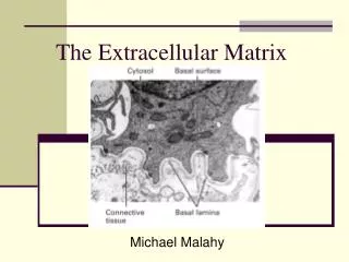
Extracellular Matrix Introduction Cell walls: bacteria, fungi, algae, and higher plants Animal cell in tissues embedded in an extracellular matrix of proteins and polysaccharides Function Provides structural support to cells and tissues Important role in regulating cell behavior Cell to cell interaction, communication
General Structure of Extracellular Matrix Animal cells embedded in an extracellular matrix Basal laminae:thin layer on which epithelial cells rest. Also surrounds muscle cells, adipose cells, and peripheral nerves most abundant in connective tissues Connective tissue loose connective tissue Bone tendon cartilage
Composition of Extracellular Matrix Fibrous proteins Polysaccharides- gel like environment Adhesion proteins- link components of the matrix to one another and to cells Different matrices have different amounts of each component Tendons, rich in fibrous proteins Cartilage, high in polysaccharides Bone, calcium phosphate crystal deposition
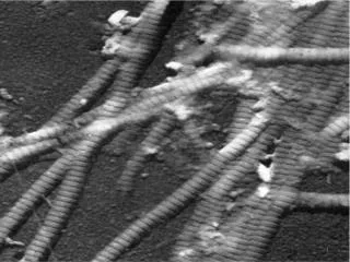
Collagen-major structural fibrous protein Forms triple helices Triple helix domains: repeats of the amino acid sequence Gly-X-Y Glycine in every 3rd position X=Pro, packs helices closely Y= hydroxyproline,synthesized in ER Pro, Hpro stabilizesby helping H-bonding Matrix composition: Collagen
Type I collagen- the most abundant polypeptide chains have about 330 Gly-X-Y repeats Secreted through ER/golgi, form collagen fibrils Triple helical molecules are associated in regular staggered arrays Covalent cross-links: lysine and hydroxylysine side chains strengthen the fibrils Fibrils form collagen fibers, several µm in diameter Matrix composition: Collagen
Some are not fibril forming Fibril-associated collagens: bind to collagen fibrils, link to others or to other matrix components Network-forming collagens: have non helical interruption, cross-link to network Anchoring fibrils: link basal laminae to underlying connective tissues Transmembrane collagens: proteins that participate in cell-matrix interactions Types of Collagen Network-forming collagens
Extracellular matrix gels are polysaccharides called glycosaminoglycans(GAGs). GAGs are repeating units of disaccharides: One sugar is either N-acetylglucosamine or N-acetylgalactosamine, the second is usually acidic (glucuronic acid or iduronic acid). Matrix Polysaccharides
sulfate groups make GAGs negatively charged bind positively charged ions and trap water molecules to form hydrated gel GAGs are linked to proteins to form proteoglycans Matrix Polysaccharides
Link matrix components to each other to cell surfaces Fibronectin : main adhesion protein of connective tissues A homodimeric protein (2500 aa/subunit), binds collagen and GAGs cells Recognized by cell surface receptors Attachment of cells to the extracellular matrix Matrix Adhesion proteins: Fibronectin
Laminin: adehsion protein of basal laminae Heterotrimeric: α, β, and γ-chains(5, 4, 3 genes, respectively) have binding sites for cell surface receptors, eg integrins type IV collagen Proteoglycans Assemble to cross-linked network Linking cells and matrix Matrix Adhesion proteins: Laminins
Cell-Matrix Interactions Integrins: major cell surface receptors, involved in attachment of cells to the extracellular matrix Transmembrane proteins, heterodimer of α and β subunits (18α, 8β) Bind to short aa in, Collagen Fibronectin laminin also anchor the cytoskeleton to the extracellular matrix
Cell-Matrix Junctions Two types of cell-matrix junction Focal adhesions:bundles of actin filaments are anchored to βsubunits of integrins via α-actinin Vinculin via talin Assembly of focal adhesions Focal complex: small group of integrins RecruiteTalin, Vinculin, α-actinin and Formin Formin initiates actin bundles
Focal adhesions are reversible Integrins can reversibly bind matrix components change conformation between active and inactive states Inactive state: integrin heads turned close to cell surface Cell signaling extends heads to matrix Migrating cells: focal adhesions form at the leading edge
Cell-Matrix Junctions: Hemidesmosomes Hemidesmosomesanchor epithelial cells to the basal lamina α6β4 integrinsbind to lamins long cytoplasmic tail of β subunit binds to intermediate filaments via Plectin and BP230 and BP180 (similar to transmembrane collagens)
Cell-Cell interactions Interactions between cells are critical for development and function of multicellular organisms Cell-cell interactions: Transient: activation of immune cells; migration to injury site Stable: role in the organization of tissues. Cell-Cell junctions allow rapid communication between cells During embryo development, cells from one tissue specifically adhere to cells of the same tissue rather than cells of a different tissue
Cell-Cell interactions Cell-cell adhesion- mediated by four groups of cell adhesion molecules Selectins, integrins, the immunoglobulin (Ig) superfamily, and cadherins Many adhesions are divalent cation-dependent, requiring Ca2+, Mg2+ or Mn2+
Selectins Selectins- transient interactions between leukocytes and endothelial cells Leukocytes slow down, flattened, migrate from the circulation to sites of tissue inflammation initial adhesion stable adhesionsbinding of integrinsto intercellular adhesion molecules(ICAMs) on endothelial cells
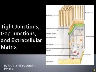
Cell to Cell Junctions Four types of Cell-Cell connections in animal cells Adherens Junctions Desmosomes Tight Junctions Gap Junctions
Adherens Junctions Cadherin form stable cell-cell connections involve actin filaments Also include β-catenin, p120, and α-catenin, β-catenin and p120 bind to cadherin and help maintain stability β-catenin binds α-catenin that interacts with actin filament of cytoskeleton
Desmosomes link the intermediate filament of adjacent cells Desmoglein and desmocollin (transmembrane cadherins) bind by heterophilic interactions across the junction Plakoglobin and plakophilin bind to the cadherins and link to the intermediate filament binding protein, desmoplakin
Tight Junctions Tight junctions provide minimal adhesive strength between the cells, usually associated with adherens junctions and desmosomes in a junctional complex
Tight Junctions Tight junctions in epithelial cell form a seal that prevents free passage of molecules and ions between cells separate apical and basolateral domains of the plasma membrane prevent free diffusion of lipids and membrane proteins
Tight Junctions transmembrane proteins, occludin, claudin, and junctional adhesion molecule (JAM), anchored on F-actin Bind similar proteins on the adjacent cell Sealing the space between cells
Gap Junctions open channels through the plasma membrane allowing ions and small molecules to diffuse freely Proteins and nucleic acids can not pass through heart muscle cells, passage of ions through gap junctions synchronizes the contractions of neighboring cells allow passage of some signaling molecules, such as cAMP and Ca2+, coordinating responses of cells in tissues
Gap Junctions Gap junctions are made of transmembrane proteins in the connexinfamily 6 connexins form a cylinder with an open aqueous pore in its center, called a connexon Connexons in the plasma membrane adjacent cells align form open channels between the two cytoplasms
Gap Junctions Specialized gap junctions occur on specific nerve cells and form an electrical synapse Individual connexons can be opened or closed When open, they allow rapid passage of ions between the two nerve cells