Download
1 / 46
610 likes | 1.65k Vues
Biochemistry of Extracellular Matrix. Jana Novotná. Composition of Extracellular Matrix (ECM). Cells (mesenchymal origin) - fibroblasts - smooth muscle cells - chondroblasts - osteoblasts and epitelial cells Organic fibrillar matrix Organic nonfibrillar matrix Water.

E N D
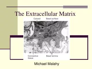
Biochemistry of Extracellular Matrix Jana Novotná
Composition of Extracellular Matrix (ECM) • Cells (mesenchymal origin) - fibroblasts - smooth muscle cells - chondroblasts - osteoblasts and epitelial cells • Organic fibrillar matrix • Organic nonfibrillar matrix • Water
Function of ECM • Provides support and anchorage for cells. • Regulates and determine cells dynamic behaviour : - polarity of cells - cell differentiation - adhesion - migration • Provides mechanical support for tissues and organ architecture - growth - regenerative and healing processes - determination and maintenance of the structure • Place for active exchange of different metabolites, ions, water.
Structure of ECM • collagen – the main ECM component, forms the main fibres • elastin • proteoglycans - heteropolysacharides • structural glycoproteins - fibronectin, laminin
Collagen • The most abundant protein in the body, making 25%-35% of all the whole-body proteins. • Collagen contributes to the stability of tissues and organs. • It maintains their structural integrity. • It has great tensile strenght. • The main component of fascia, cartilage, ligaments, tendons, bone and skin. • Plays an important role in cell differentiation, polarity, movement. • Plays an important role in tissue and organ development.
Collagen Structure • Collagen is insoluble glycoprotein (protein + carbohydrate) • Collagen polypeptide primary structure: • -G – X – A – G – A – A – G – Y – A – G – A – A – G – X – A – G− • , • G - glycine, X - proline or hydroxyproline, Y – lysin or hydroxylysine, A – amino acid • Proline and hydroxyproline constitute about 1/6 of the total sequence,provide the stifness of the polypeptide chain. • Carbohydrates : glucose, galactose
Three helical polypeptide units twist to form a triple-helical collagen molecule: a molecular "rope" which has some bending stiffness and does not undergo rotation. • The tropocollagen molecule has a length of approximately 300 nm and a diameter close to 1.5 nm. • In the typical fibrillar collagens, only short terminal portions of the polypeptides (the telopeptides) are not triple helical.
Synthesis • Synthesis of a chainsof pre-procollagen on ribosomes. • Hydroxylationof lysine and proline in rER/Golgi by lysyl-5-hydroxylase and prolyl-4-hydroxylase. • Glycosylation: addition of galactose and glucose to some hydroxylysine residues (galactosyl transferase and glycosyl transferase). • Assembly of a-chains to form procollagen. Reaction needs the formation of disulphide bonds between registration peptides, at both ends of theprepro- collagen.
5. Secretion of procollagen molecules by exocytosis into theextracellular space. 6. Cleavage of registration peptides is catalysed by procollagen peptidases.The resulting molecule is called tropocollagen. 7. Oxidation – deamination of the hydroxylysine, the removal of (NH2) group has a net oxidative effect and the formation of covalent cross-links. Reaction is catalyzed by lysine oxidase (or catalase). 8. Self-assembly or polymerization of tropocollagen molecules form collagen fibrils.Cross-linkage between adjacent tropocollagen molecules stabilizes the fibrils.
Posttranslation Modification of Collagen Molecule Hydroxylation of some prolyl and lysyl residues prolyl-4-hydroxylase, lysyl-5-hydroxylase Cofactors: O2 (or superoxid) a-ketoglutarate Fe2+ vitamin C (ascorbic acid) Proline + a-ketoglutarate+ O2 + Fe2+ → 4-hydroxy-proline + Fe3+ + CO2 + succinate Hydroxyproline stabilizes molecule oftropocollagen.
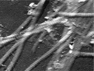
The typical staggered array of tropocollagen molecules in the collagen fibril. The telopeptides participate in covalent crosslinking.
Collagen – Fiber Formation Collagen types I, II, III, V, IX, X Cross striated structure of collagen fiber reflect periodic composition of individual tropocollagen molecules. Collagen fibrils of 1 mm diameter support the weight of 10 kg.
Collagen Interactions Fiber forming collagen and nonfibrous collagen Cartilagous matrix Tendon
Collagens Classification • Fibril-forming collagens (I, II, III, V, X) • Fibril-associated collagens (FACIT) • Network-forming collagens • Anchoring fibrils collagens • Transmembrane collagens • Basement membrane collagens • Other collagens with unique function
Major Collagen Types Fibril forming collagens (Most abundant collagen family - 90 % of the total collagens)
Basement membrane collagens Short non-helical amino-terminal domain, a long Gly-X-Y repeat domain with numerous small interruptions, and a highly conserved carboxyl-terminal globular NC1 domain. It polymerizes into a disulfide-bonded polygonal network via tetramerization between amino-terminal domains and dimerization between NC1 domains.
Elastin • Elastin is a major protein component of tissues that require elasticity such as arteries, lungs, bladder, skin and elastic ligaments and cartilage. • It is composed of soluble tropoelastin protein containing primarily glycine and valine and modified alanine and proline residues. • Tropoelastin is a ~65kDa protein that is highly cross-linked to form an insoluble complex. • Polypeptide chains are cross-linked together to form rubberlike, elastic fibers. Each elastin molecule uncoils into a more extended conformation when the fiber is stretched and will recoil spontaneously as soon as the stretching force is relaxed. Jaime Moore , Susan Thibeault Journal of Voice Volume 26, Issue 3 2012 269 - 275
Elastin • Desmosine (isodesmosine) - the most common interchain cross-link • conversion of NH3 groups of lysine (hydroxylisine) to reactive aldehydes by lysyl oxidase. • desmosine cross-link isspontaneously formed.
Proteoglycans Special class of glycoproteins heavily glycosylated (95%). Core protein with one or more attached glycosamino glycan chain(s).
Glycosaminoglycans (GAG) • Long chain, linear carbohydrate polymers • Negatively charged under physiological conditions (due to the occurrence of sulfate and uronic acid groups). Disaccharide subunits are: 1. uronic acid D-glucuronic acid or L-iduronic acid 2. aminosugar N-acetyl glucosamin (GlcNAc) or N-acetyl galactosamin (GalNAc)
Amino sugars and uronic acids are the most common building blocks of the glycosaminoglycans. • amino sugars -OH at C-2 is replaced by an amino group. This amino group is most often acetylated and sometimes sulfated. • uronic acidsC-6 of the hexose is oxidized to a carboxyl group.
Linkage of GAGs to protein core by specific trisaccharide linker
Biosynthesis • The protein component is synthesized by ribosomes and transocated into the lumen of the RER. • Glycosylation of the proteoglycan occurs in the Golgi apparatus in multiple enzymatic steps. • First a special link tetrasaccharide is attached to a serine side chain on the core protein to serve as a primer for polysaccharide growth.
Biosynthesis • Then sugars are added by glycosyltransferase. • Some glycosyltransferases catalyse sugar transfer to tyrosine, serine and threonine to give O-linked glycoproteins, or to asparagine to give N-linked glycoproteins. • Mannosyl groups may be transferred to tryptophan to generate C-manosyl tryptophan • The completed proteoglycan is then exported in secretoryvesicles to the extracellular matrix of the cell.
Glycosaminoglycan Occurence Proteoglycans can be categorised depending upon the nature of their glycosaminoglycan chains. • Hyaluronic acid (does not contain any sulfate) • non-covalent link complex with proteoglycans • Chondroitinsulfate • cartilage, bone • Dermatan sulfate • skin, blood vessels • Heparan sulfate • basement membrane, component of cells surface • Keratan sulfate • cornea, bone, cartilage, often aggregated with chondroitin sulfate
Function of Proteoglycans • organize water molecules - resistent to compression - return to original shape - repel negative molecules • occupy space between cells and collagen • high viscosity - lubricating fluid in the joints • specific binding to other macromolecules • link to collagen fibers - form network - in bone combine with calcium salts (calcium carbonate,hydroxyapatite) • cell migration and adhesion - passageways between cells • anchoring cells to matrix fibers
Structural Glycoproteins • Direct linkage to collagen or proteoglycans • anchoring collagen fibers to cell membrane • covalent attachment to membrane lipid • Major adhesive structural glycoproteins • fibronectin • laminin
Fibronectin • High-molecular weight (~440kDa) glycoprotein • Attached to cell membrane by membrane-spanning receptor – integrin. • Crosslinks and stabilizes other components of ECM • Enhances cell addhesion to extracellular matrix components (collagen, fibrin and heparansulfate proteoglycans). • Related to blood clotting - soluble FN crosslinks platelets together using membrane bound heparin
FibronectinStructure • protein dimer connected at C-terminal by S-S linkage • rigid and flexible domains • cell binding domain RGD • (Arg, Gly, Asp) • - binding receptor in cell membranes • RGD domain binds to • - collagen type I, II and III • - heparin sulfate • - hyaluronic acid • - fibrin
Fibronectin Function • related to cell adhesion, differentiation, growth, migration; • anchoring basal laminae to other ECM; • plasma fibronectin forms a blood cloth, along with fibrin; • related to cell movement - groups of embryonic cells follow a FN pathway -FN guides macrophages into wound areas.
Laminin structure • and function • cross-shapedglycoprotein • 3 polypeptide chains • domain bind • - collagen type IV • - heparin • - heparin sulfate • cell surface receptor RGD • cell adhesion • role in cell differentiation • anchoring the glycoprotein to basal laminae
Fibrillin • Glycoprotein, essential for formation of elastic fibers(asheath surrounding the amorphous elastin) • Produced by fibroblasts. • A group of three proteins, fibrillin-1, -2 and -3. • The main role - maintaining the structural integrity of tissues, the regulation of cytokines – TGF-b • In humans, defects in the fibrillin-1 and fibrillin-2 genes have been linked to diseases that affect the cardiovascular, skeletal and ocular systems, including Marfan syndrome.
Integrins • Groups of transmembrane protein receptors • mediate the attachment between a cell and its surroundings • integrins perform outside-in signaling and they also operate an inside-out mode. • Link cytoskeleton to ECM • Fibronectin receptor is best known
Tanescins • Abundant in the extracellular matrix of developing vertebrate embryo. • Tenascin-C contains an RGD motif and is recognized by diverse integrins. Mainly synthesized by the nervous system and connective tissues. • Tenascin-R is found in the nervous system • Tenascin-X and tenascin-Y are found primarily in muscle connective tissues. • Tenascin-W is found in kidney and developing bone.