Download
1 / 20
310 likes | 1.43k Vues
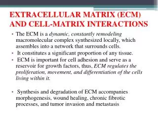
EXTRACELLULAR MATRIX PROTEINS AND PROTEINASES. By, Raghu Ambekar. Photonics Research of Bio/nano Environments Department of Electrical & Computer Engineering University of Illinois Urbana - Champaign. BioE 506. Outline. Extracellular matrix proteins Collagen Classification

E N D
EXTRACELLULAR MATRIX PROTEINS AND PROTEINASES By, Raghu Ambekar Photonics Research of Bio/nano Environments Department of Electrical & Computer Engineering University of Illinois Urbana - Champaign BioE 506
Outline • Extracellular matrix proteins • Collagen • Classification • Fibril assembly and collagen diseases • Extracellular matrix proteinases • Role of MMP in metastasis • Modification of tumor collagen for therapeutics
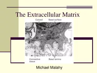
Extracellular matrix (ECM) • Surrounds cell • Provides mechanical support • Controls the flow of nutrients and signals to the cells • Consists of • Fibrous: collagen, elastin, fibronectin, laminin • Non-fibrous: Proteoglycans and polysaccharides http://kentsimmons.uwinnipeg.ca/cm1504
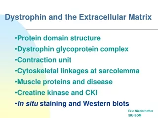
Collagen • Collagen : most abundant protein found in the human body. About 1/3rd of the total proteins. • Found abundantly in tendon, cartilage, bone and skin • Functions: • cell migration • cell adhesion • molecular filtration • tissue repair
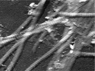
Structure of collagen • It has a triple-helix structure containing three α-polypeptide chains arranged in right-handed supercoil • Glycine, proline, hydroxyproline • 1.5 nm diameter • At least 28 different collagens found • The three α-chains could be same (collagen II) or different (collagen I) Collagen molecule
Classification of collagen 1. Fibril-forming collagens • No interruptions in triple helix • Regular arrangement results in characteristic “D” period of 67 nm • Diameter : 50-500 nm • Example : Types I, II, III, V, XI
Classification of collagen 2. Network-forming collagens • Forms network in basement (Collagen IV) and Descemet’s membrane (Collagen VIII) • Molecular filtration • Example : Types IV, VIII, X
Classification of collagen 3. Fibril-associated collagens with interrupted triple helices (FACITs) • Short collagens with interruptions • Linked to collagen II and carries a GAG chain • Found at the surface of fibril-forming collagens • Example : Types IX, XII, XIV
Classification of collagen 4. Anchoring collagens • Provides functional integrity by connecting epithelium to stroma • Example : Type VII
Classification of collagen 5. Beaded-filament-forming collagens • Form structural links with cells • Example : Type VI • Collagen VI crosslink into tetramers that assemble into long molecular chains (microfibrils) and have beaded repeat of 105 nm
Type I Fibril assembly Fibril assembly is determined by chain recognition sequence in C-propeptide Fish scale Bone osteon Tendon Chain recognition sequence Skin
Diseases associated with collagen • Diseases caused by mutations • Subtypes of osteogenesis imperfecta (collagen I) • Ehlers-Danlos syndrome (collagen I and V) • Alport syndrome (collagen IV) • Certain arterial aneurysms (collagen III) • Ullrich muscular dystrophy (collagen VI) • Certain chondrodysplasias (collagen IX and XI) • Kniest dysplasia (collagen II)
Role of MMP in metastasis Metastasis • Metastasis • Spread of cancer from a primary tumor to distant sites of the body • A defining feature of cancer
Role of MMP in metastasis • Understanding the molecular mechanisms of metastasis is crucial for the design of therapeutics • Extracellular matrix metalloproteinases (MMP) associated with metastasis • MMPs are capable of digesting ECM and basement membrane under physiologic conditions • Collagenases degrade fibrillar collagen • Stromelysins degrade proteoglycans and glycoproteins • Gelatinases degrade nonfibrillar and denatured collagens • At tumor sites, experiments have found • Increased number of MMPs • Increased levels of MMPs • Reduced levels of TIMPs (Tissue inhibitors of metalloproteinases)
Role of MMP in metastasis • Major role of MMPs was to facilitate the breakdown of physical barriers, thus promoting invasion, intravasation, extravasation and migration • MMPs targeted for antimetastasis therapies
Role of MMP in metastasis • Clinical trials of inhibiting MMPs to cure cancer have failed • Metastasis is a complicated process • MMPs contribute to every stage in tumor progression at both primary and metastatic sites • Specific MMPs play a role in each stage of metastasis • MMP 13, 14 – invasion • MMP 9– angiogenesis • Understand the role of the MMPs in each cancer setting
Modification of collagen for therapeutics • Structure and content of collagen governs the delivery of therapeutic molecules in tumors • Penetration of therapeutic molecules improved by developing agents that modify ECM and increase diffusion • Detect tumor collagen noninvasively to quantify collagen content and estimate drug delivery characteristics
Modification of collagen for therapeutics Uses Second-harmonic generation (SHG) for imaging only collagen fibers SHG: Blue Wavelength=400 nm Red Wavelength=800 nm • Conditions : • Non-centrosymmetric (collagen, microtubules) • Lasers (high intensity) SAMPLE Collagen stained red and imaged by fluorescence microscopy • Advantages : • No staining • 3D imaging • No photobleaching Collagen imaged by SHG microscopy
Modification of collagen for therapeutics • SHG intensity collected from live imaging of collagen fibers provides an good estimate of diffusion coefficient in tumors
Modification of collagen for therapeutics 0th day 3rd day 6th day 9th day 12th day • Chronic relaxin treatment degrades tumor matrix and improve macromolecular diffusion in tumors THANK YOU!