Quantitative Physiology
Quantitative Physiology. Module (2). Cardiovascular System. Amr A. Sharawi. Cardiovascular System – The Intended Function. Blood flows through organs and tissues either to nourish and cleanse them or to be itself processed in some sense, e.g.; oxygenated (pulmonary circulation)

Quantitative Physiology
E N D
Presentation Transcript
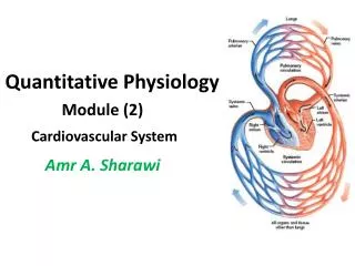
Quantitative Physiology Module (2) Cardiovascular System • Amr A. Sharawi
Cardiovascular System – The Intended Function • Blood flows through organs and tissues either to nourish and cleanse them or to be itself processed in some sense, e.g.; • oxygenated (pulmonary circulation) • stocked with nutrients (splanchnic or portal circulation) • filtered of used red blood cells (splenic circulation) • dialyzed (renal circulation) • cooled (cutaneous circulation)
Arrangement of the parallel routes by which the circulation passes from the aorta to the vena cava.
PATHWAYS OF CIRCULATION • PULMONARY CIRCULATION • It begins at the right ventricle, continues through the lungs, and terminates at the left atrium. • SYSTEMIC CIRCULATION • It begins at the left ventricle, continues through all other body systems in parallel pathways, and terminates at the right atrium. • HEPATIC PORTAL (SPALANCHNIC) CIRCULATION • This is a subdivision of systemic circulation in which blood from the abdominal digestive organs and spleen circulates through the liver before returning to the heart.
Splanchnic Circulation • The splanchnic circulation is composed of a parallel network of the following circulations: • gastric • small intestinal • colonic • pancreatic • hepatic • splenic
Simplified schematic of splanchnic vascular bed showing the parallel pathways of the circulationof the various gastrointestinal organs and their series arrangement with the portal circulation tothe liver, and the common venous drainage of all these organs.
The Vascular System - A Highway Network • Every cell in the human body is near enough to the environment to easily exchange with it: • mass (including nutrients, oxygen, carbon dioxide, and the waste products of metabolism) • energy (including heat) • The human body is endowed with a major highway network — organized to make available thousands of miles of access tubing for the transport to and from a different neighborhood of any given cell whatever it needs to sustain life whatever it needs to sustain life.
The Vascular System - A Highway Network • This highway network, called the cardiovascular system, cardiovascular system, includes: • a pumping station, the heart; • a working fluid, blood; • a complex branching configuration of distributing and collecting pipes and channels, blood vessels; • a sophisticated means for both intrinsic (inherent) and extrinsic (autonomic and endocrine) control.
Path of blood flow through the entire cardiovascular system. All the structures within the colored box are located in the heart.
The Working Fluid: Blood • Blood is a complex, heterogeneous suspension of formed elements, the blood cells, or hematocytes, suspended in a continuous fluid called plasma. It is further • Accounting for about 8 ± 1% of total body weight • Averaging in volume 5200 ml • Nominally, the composite fluid has a mass density of 1.057 ± 0.007 g/cm3 and it is 3 to 6 times as viscous as water.
Measurement of the hematocrit by centrifugation. Due to the presence of a thin layer of leukocytes and platelets between the plasma and red cells, the value for plasma determined bycentrifugation is actually slightly less than 55 percent.
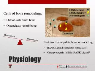
Hematocytes • Hematocytes are all derived in the active (“red”) bone marrow of adults from undifferentiated stem cells called hemocytoblasts, and all reach ultimate maturity via a process called hematocytopoiesis. • They include 3 basic types of cells: • red blood cells (erythrocytes, totaling nearly 95% of the formed elements) • white blood cells (leukocytes, averaging <0.15% of all hematocytes) • platelets (thrombocytes, on the order of 5% of all blood cells)
Hematocytes • The primary function of erythrocytes is to aid in the transport of blood gases. • The primary function of leukocytes is to provide the human body with the ability to identify and dispose of foreign substances such as infectious organisms that do not belong there. • The primary function of platelets is to participate in the blood clotting process. • Removal of all hematocytes from blood by centrifugation or other separating techniques leaves behind the aqueous saline suspending medium called plasma.
Plasma • Plasma has an average mass density of 1.035 ± 0.005 g/cm3 and a viscosity 1½ to 2 times that of water and its almost 91% water by weight. • Plasma contains plasma proteins, of which there are three major types: • Albumin • Globulins • fibrinogen • Other plasma constituents are minerals, trace elements, and electrolytes, mostly cations: sodium, potassium, calcium, and magnesium; anions: chlorine, bicarbonate, phosphate, and sulfate
Plasma Proteins • Primary functions of albumin: • help maintain the osmotic transmural pressure differential that ensures proper mass exchange between blood and interstitial fluid at the capillary level. • serve as a transport carrier molecule for several hormones and other small biochemical constituents (such as some metal ions). • Primary functions of the globulins: • act as transport carrier molecules for large biochemical substances, such as fats and certain carbohydrates and heavy metals. • work together with leukocytes in the body’s immune system. • Primary function of fibrinogen: • work with thrombocytes in the formation of a blood clot—a process also aided by one of the most abundant of the lesser proteins, prothrombin.
Serum • Removal from blood of all hematocytes and the protein fibrinogen (by allowing the fluid to completely clot before centrifuging) leaves behind a clear fluid called serum, which has a density of about 1.018 ± 0.003 g/cm3 and a viscosity up to 1½ times that of water.
Laminar and Turbulent Flow of Blood in Vessels: • When blood flows at a steady rate through a long, smooth blood vessel, it flows in streamlines, with each layer of blood remaining vessel, at the same distance from the vessel wall. • Also, the central most portion of the blood stays in the center of the vessel. • When laminar flow occurs, the velocity of flow in the center of the vessel is far greater than that toward the outer edges. This effect is called the “parabolic profile for velocity of blood flow”. • In turbulent flow, blood is flowing in all directions in the vessel and continually mixing within the vessel.
Turbulent Flow • Some reasons for turbulent flow of blood are: • when the rate of blood flow becomes too great • when it passes by an obstruction in a vessel • when it makes a sharp turn • when it passes over a rough surface • In these cases the flow may then become turbulent, or disorderly, rather than streamline. • Turbulent flow means that the blood flows crosswise in the vessel as well as along the vessel, usually forming whirls in the blood called eddy currents vessel. • These are similar to the whirlpools that one frequently seen in a rapidly flowing river at a point of obstruction. • When eddy currents are present, the blood flows with much greater resistance than when the flow is streamline because eddies add tremendously to the overall friction of flow in the vessel.
Diagrammatic representation of normal laminar flow in comparison with turbulent flow
Example of normal laminar flow through aortic valve (top) and turbulent flow resulting from aortic stenosis(bottom)
Reynolds’ Number • The tendency for turbulent flow increases in direct proportion to the velocity of blood flow v, the diameter of the blood vessel d, and the density of the blood ρ, and is inversely proportional to the viscosity of the blood η, in accordance with the following equation: • Re is Reynolds Re is Reynolds’’ number and is the measure of the tendency for turbulence to occur.
Reynolds’ Number • When Reynolds’ number rises above 200 to 400, turbulent flow will occur at some branches of vessels but will die out along the smooth portions of the vessels. • However, when Reynolds’ number rises above approximately 2000, turbulence will usually occur even in a straight, smooth vessel. • Reynolds’ number for flow in the vascular system even normally rises to 200 to 400 in large arteries; as a result there is almost always some turbulence of flow at the branches of these vessels. • In the proximal portions of the aorta and pulmonary artery, Reynolds’ number can rise to several thousand during the rapid phase of ejection by the ventricles; this causes considerable turbulence in the proximal aorta and pulmonary artery where many conditions are appropriate for turbulence: • (1) high velocity of blood flow • (2) pulsatile nature of the flow • (3) sudden change in vessel diameter • (4) large vessel diameter
The Pumping Station: The Heart • Barely the size of the clenched fist of the individual in whom it resides—an inverted, conically shaped, hollow muscular organ measuring 12 to 13 cm from base (top) to apex (bottom) and 7 to 8 cm at its widest point and weighing about 325 g, the human heart occupies a small region between the third and sixth ribs in the central portion of the thoracic cavity of the body. • It rests on the diaphragm, between the lower part of the two lungs, its base-to-apex axis leaning mostly toward the left side of the body and slightly forward. • The heart is divided by a tough muscular wall, the interatrial-interventricular septum, into a right side and a left side, each being one self-contained pumping station, but the two being connected in series.
Diagrammatic section of the heart. The arrows indicate the direction of blood flow.
The Two Sides of the Heart • The left side of the heart drives oxygen-rich blood through the aortic semilunar outlet valve into the systemic circulation, which carries the fluid to within a differential neighborhood of each cell in the body—from which it returns to the right side of the heart low in oxygen and rich in carbon dioxide. • The right side of the heart then drives this oxygen-poor blood through the pulmonary semilunar outlet valve into the pulmonary circulation, which carries the fluid to the lungs, where its oxygen supply is replenished and its carbon dioxide content is eliminated before it returns to the left side of the heart to begin the cycle all over again. • Because of the anatomic proximity of the heart to the lungs, the right side of the heart does not have to work very hard to drive blood through the pulmonary circulation, so it functions as a low-pressure (P≤ 40 mmHg gauge) pump compared with the left side of the heart, which does most of its work at a high pressure (up to 140 mmHg gauge or more) to drive blood through the entire systemic circulation to the furthest extremes of body.
The Heart Chambers • Each cardiac pump is further divided into two chambers: a small upper receiving chamber, or atrium, separated by a one-way valve from a lower discharging chamber, or ventricle, which is about twice the size of its corresponding atrium. • Altogether, the heart chambers collectively have a capacity of some 325 to 350 ml, or about 6.5% of the total blood volume in a “typical” individual—but these values are not actual, since the organ alternately fills and expands, contracts, and then empties as it generates a cardiac output.
Phases of the Heart Cycle • Filling phase (diastole) • Emptying phase (systole)
Diastole • It takes 480-ms or so of the average 750-ms cardiac cycle. During this phase: • The inlet valves of the two ventricles are open. These are: • the 3.8-cm-diameter tricuspid valve from right atrium to right ventricle • the 3.1-cm diameter bicuspid or mitral valve from left atrium to left ventricle • The outlet valves are closed. These are: • the 2.4-cm diameter pulmonary valve • the 2.25-cm-diameter aortic semi-lunar valve • The heart ultimately expands to its end-diastolic volume (EDV), which is on the order of 140 ml of blood for the left ventricle.
Systole • During the 270-ms emptying phase (systole) electrically induced vigorous contraction of cardiac muscle drives the intraventricular pressure up, thus forcing: • the one-way inlet valves closed • the unidirectional outlet valves open • The heart contracts to its end-systolic-volume (ESV), which is typically on the order of 70 ml of blood for the left ventricle. • The ventricles normally empty about half their contained volume with each heart beat, the remainder being termed the cardiac reserve volume.
Photographs of the pulmonary valve from the pulmonary trunk looking down into the right ventricle. On the top the valve is in the process of opening as blood flows through it from the right ventricle into the pulmonary trunk. On the bottom, the valve is in the process of closing, the cusps being forced together when the pressure of the blood in the pulmonary trunk is greater than the pressure in the right ventricle.
Summary of events in the left atrium, left ventricle, and aorta during the cardiac cycle.
Stroke Volume • The difference between the actual EDV and the actual ESV, called the stroke volume (SV). • This is the volume of blood expelled from the heart during each systolic interval • The ratio of SV to EDV is called the cardiac ejection fraction, or ejection ratio: • 0.5 to 0.75 is normal, • 0.4 to 0.5 signifies mild cardiac damage, • 0.25 to 0.40 implies moderate heart damage, • less than 0.25 warns of severe damage to the heart’s pumping ability)
Cardiac Output • If the stroke volume is multiplied by the number of systolic intervals per minute, or heart (HR), one obtains the total cardiac output (CO): CO = HR × (EDV – ESV) • Dawson [1991] has suggested that the cardiac output (in milliliters per minute) is proportional to the Weight W (in kilograms) of an individual according to the equation CO = 224W3/4 • and that “normal” heart rate obeys very closely the relation HR = 229W(-1/4)
Example • For a “typical” 68.7-kg individual (blood volume = 5200 ml) the previous equations yield: • CO = 5345 ml/min, • HR = 80 beats/min • SV = CO/HR = 67.2 ml/beat, which are very reasonable values. • Furthermore, assuming this individual lives about 75 years, his or her heart will have cycled over 3.1536 billion times, pumping a total of 210.7 million liters of blood (7692 liters per day)—all of it emptying into the circulatory pathways that constitute the vascular system.
The Piping Network: Blood Vessels • The vascular system is divided by a microscopic capillary network into: • a downstream upstream, high-pressure, arterial side consisting of relatively thick-walled, viscoelastic tubes that carry blood away from the heart • an upstream, low-pressure, venous side consisting of correspondingly thinner (but having a larger caliber) elastic conduits that return blood back to the heart.
Normal blood pressures in the different portions of the circulatory system when a person is lying in the horizontal position.
Vascular Wall • Except for their differences in thickness, the walls of the largest arteries and veins consist of the same three distinct, well-defined, and well developed layers. • the thinnest tunica intima • the thickest tunica media composed of: • numerous circularly arranged elastic fibers • a significant amount of smooth muscle cells arranged in spiraling layers around the vessel wall • some interlacing collagenous connective tissue • the medium-sized tunica adventitia
Blood vessel structure is directly related to function: A, Demonstration of the effect of vessel diameter on blood flow. B, Concentric rings of blood flowing at different velocities; the farther away from the vessel wall, the faster the flow.
Poiseuille’s Law • The concentric rings inside the vessels indicate that the velocity of flow in each ring is different from that in the adjacent rings because of laminar flow. • Blood in the ring touching the wall of the vessel is barely flowing, because of its adherence to the vascular endothelium. • The next ring of blood toward the center of the vessel slips past the first ring and, therefore, flows more rapidly. • The third, … rings likewise flow at progressively increasing velocities. • Thus, the blood that is near the wall of the vessel flows extremely slowly, whereas that in the middle of the vessel flows extremely rapidly. • In the small vessel, essentially all the blood is near the wall, so that the so that the extremely rapidly flowing central stream of blood simply does not exist.
Poiseuille’s Law • Poiseuille’s law is given by: In other words: • where R = blood flow resistance
Blood Flow Resistance • In the systemic circulation, the most resistance to blood flow is small arteriolar resistance. • The internal diameters of the arterioles range from as little as 4 μm to as great as 25 μm. • However, their strong vascular walls allow the internal diameters to change tremendously, often as much as fourfold. • From the fourth power law that relates blood flow to diameter of the vessel, one can see that a fourfold increase in vessel diameter can increase the flow as much as 256-fold. • Thus, this fourth power law makes it possible for the arterioles, responding with only small changes in diameter to nervous signals or local tissue chemical signals, either to turn off almost completely the blood flow to the tissue or at the other extreme to cause a vast increase in flow.
Vascular resistances: A, in series and B, in parallel.For A:For B:
Electrical Analogy of the Cardiovascular System • In a global sense, one can think of the human cardiovascular system using an electrical analogy as: • a voltage source (the heart), • two capacitors (a large venous system and a smaller arterial system), and • a resistor (the microcirculation taken as a whole). • The cardiovascular system is designed to bring blood to within a capillary size of each and every one of the more than 1014 cells of the body—but which cells receive blood at any given time, how much blood they get, the composition of the fluid coursing by them, and related physiologic considerations are all matters that are not left up to chance.