Physiology
Physiology. Skeletal Muscular Contraction. http://www.youtube.com/watch?v=uRoRsmvkhTI&eurl=http://wendyusuallywanders.wordpress.com/?tag=myasthenia-gravis&feature=player_embedded. Connective Tissue. Endomysium Surrounds each muscle fiber (cell) Attaches to Z-lines in each sarcomere

Physiology
E N D
Presentation Transcript
Physiology Skeletal Muscular Contraction
http://www.youtube.com/watch?v=uRoRsmvkhTI&eurl=http://wendyusuallywanders.wordpress.com/?tag=myasthenia-gravis&feature=player_embeddedhttp://www.youtube.com/watch?v=uRoRsmvkhTI&eurl=http://wendyusuallywanders.wordpress.com/?tag=myasthenia-gravis&feature=player_embedded
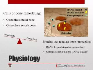
Connective Tissue • Endomysium • Surrounds each muscle fiber (cell) • Attaches to Z-lines in each sarcomere • Perimysium • Surrounds bundles (fascicles) of muscle fibers • Attaches to endomysium • Epimysium • Attaches to the Perimysium • Continuous with tendon
Sarcomere • Repeating Patterns within the myofibrils • Myofibrils • Proteins within the myofibers • Myosin • Actin
Muscle Anatomy • Sarcolemma • Muscle fiber cell membrane • Myofibrils • Highly organized bundles of contractile and elastic proteins • Carries out the work of contraction
Myofibrils = Contractile Organelles of Myofiber Contain 6 types of protein: • Actin • Myosin • Tropomyosin • Troponin • Titin • Nebulin Contractile Regulatory Accessory
Titin and Nebulin • Titin: biggest protein known (25,000 aa); elastic! • Stabilizes position of contractile filaments • Return to relaxed location • Nebulin: inelastic giant protein • Alignment of A & M
Myosin • Myo- muscle • Motor protein of the myofibril • Thick filament • Attaches to the M-line • Heads point towards Z-lines • Myosin heads are clustered at the ends of the filament • Myosin tails are bundled together
Actin • Thin Filament • Attached to Z-lines • Globular protein • G-Actin • Has binding site for myosin head • Forms a Cross-Bridge when myosin binds to G-actin • Five Actin proteins surround the myosin in 3-D pattern
Binding sites Actin filament Strong binding Weak binding Myosin head group S2 link Stretching of the link generates tension Myosin filament
Why do thin filaments move? Net force Net force Equal and opposite force on thick filament
Actin • Tropomyosin • Protein that covers over the myosin binding site on G-Actin • Myosin head can’t bind to G-Actin, muscle relaxes • If the binding site on G-Actin is uncovered by removing Tropomyosin then myosin and actin bind, muscle contracts
Actin • Troponin C • Protein attached to Tropomyosin • When Troponin C changes shape it pulls on Tropomyosin • Calcium binding to Troponin C causes this protein to change shape • Tropomyosin moves and uncovers the binding site on G-Actin, so Actin and Myosin can bind • Contraction
Regulation of Contraction by Troponin and Tropomyosin • Tropomyosin blocks myosin binding site (weak binding possible but no powerstroke) • Troponin controls position of tropomyosin and has Ca2+ binding site • Ca2+ present: binding of A & M • Ca2+ absent: relaxation
Muscle Anatomy • Sarcoplasmic Reticulum • Modified endoplasmic reticulum • Wraps around each myofibril like a piece of lace • Stores Calcium • Terminal Cisternae • Longitudinal tubules • Transverse tubules (T-tubules) • Triad-two flanking terminal cisternae and one t-tubule • T-tubules are continuous with cell membrane
Role of calcium Tropomyosin Troponin complex • Troponin and Tropomyosin bind to actin • block the actin – myosin binding sites • Troponin is a calcium binding protein
When Troponin binds calcium it moves Tropomyosin away from the actin-myosin binding site Ca Ca
Where does Calcium come from? • Intracellular storage called Sarcoplasmic Reticulum • Surround each myofibril of the whole muscle • Contains high concentration of calcium • Transverse Tubules connects plasma membrane to deep inside muscle
T-Tubules • Rapidly moves action potentials that originate at the neuromuscular junction on the cell surface
Membrane depolarization or APs carried deep into the muscle by T-tubules Motor nerve T-tubule + Neurotransmitter receptors SR
My SR Ryanodine Receptor Dihydropyridine receptor T-tubule SR myoplasm
Ca++ Ca++ Ca++ SR Ca++ pump Myoplasm (intracellular) _ _ _ + _ + _ + + _ + + _ _ + _ + _ T-tubule (extracellular) _ + + + _ +
Sliding Filament Theory • When myosin binds to the binding site on G-actin muscular contraction occurs. • The more myosin that bind to G-actin the greater the force of contraction • Calcium must be present
Sliding Filament Theory • Cross Bridge • Myosin in the High Energy Configuration binds to G-Actin • ADP + Pi are bonded to the myosin head when the cross bridge forms • Power Stroke • When the myosin and actin bind the myosin head changes shape • Myosin pulls the actin and pulls on the Z-line • Sarcomere shortens • ADP+Pi no longer binds to myosin head
Sliding Filament Theory • ATP binds to the myosin head • Myosin changes to its Low Energy Confirmation • In the Low Energy Confirmation Myosin breaks its bonds with Actin • Rigor Mortis • Lack of ATP • Build up of Lactic Acid
Sliding Filament Theory • ATPase • ATP is hydrolyzed to ADP + Pi • ATPase is on the myosin head • Myosin changes shape back to its High Energy Confirmation
Sliding Filament Theory • Some Myosin heads detach from Actin while other heads continue to keep their attachments • No slipping of the Z-lines • Contraction is held in place
What if we don’t have this? X ATP Actin + myosin Actomyosin complex Rigor mortis
Events at Neuromuscular Junction • Converts a chemical signal from a somatic motor neuron into an electrical signal in the muscle fiber
Events at Neuromuscular Junction • Acetylcholine (Ach) is released from the somatic motor neuron • Ach initiates an action potential in the muscle fiber • The muscle action potential triggers calcium release from the sarcoplasmic reticulum • Calcium combines with troponin C and initiates contractions
Events at Neuromuscular Junction • Ach binds to cholinergic receptors on the motor end plate • Na+ channels open • Na+ influx exceeds K+ efflux across the membrane • End-Plate Potential (EPP) • EPP reaches threshold and initiates a muscle action potential
Events at Neuromuscular Junction • Action Potentials move down the membrane • K+ builds up in the t-tubules • Depolarization occurs • Calcium gates on the SR opens • Calcium diffuses into the cytoplasm of the cell
Excitation-Contraction Coupling • The process where muscle action potentials initiate calcium signals that in turn activates a contraction-relaxation cycle
Initiation of Contraction Excitation-Contraction Coupling explains how you get from AP in axon to contraction in sarcomere ACh released from somatic motor neuron at the Motor End Plate AP in sarcolemma and T-Tubules Ca2+ release from sarcoplasmic reticulum Ca2+ binds to troponin
Details of E/C Coupling Nicotinic cholinergic receptors on motor end plate = Na+ /K+ channels Net Na+ entry creates EPSP AP to T-tubules DHP (dihydropyridine) receptors in T-tubules sense depolarization