physiology
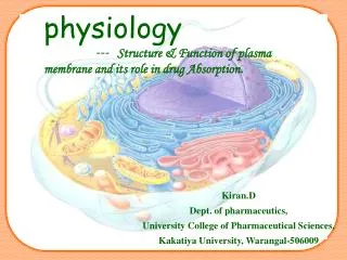
physiology --- Structure & Function of plasma membrane and its role in drug Absorption. Kiran.D Dept. of pharmaceutics, University College of Pharmaceutical Sciences, Kakatiya University, Warangal-506009. Content. Introduction Physiology of Plasma membrane. Structure,

physiology
E N D
Presentation Transcript
physiology --- Structure & Function of plasma membrane and its role in drug Absorption. Kiran.D Dept. of pharmaceutics, University College of Pharmaceutical Sciences, Kakatiya University, Warangal-506009
Content • Introduction • Physiology of Plasma membrane. • Structure, • composition, • functions. • Transport across cell membrane. • Conclusion • References
Plasma membrane structure • Definition: The Plasma membrane is a thin bi- layered structure which surrounds each cell, consists of lipids (phospholipids 75%, cholesterol 20%,glycolipids 5%), proteins (partially or completely embedded), carbohydrates etc., • ~6-10 nm thick. • Plasma membrane is asymmetrical
Membrane Models • Robertson- Unit membrane • Singer and Nicolson - Fluid-Mosaic Model • - Membrane structure is not rigid (fluid) • - Membrane comprised of diff. molecules (mosaic) • - Proteins float around the surface of the cells • - Proteins, Carbohydrates, Phospholipids can be • added/removed from the surfaces of cells
FLUID MOSAIC MODEL The fluid mosaic modeldescribes the plasma membrane as a flexible boundary of a cell. The phospholipids move within the membrane. FLUID- because individual phospholipids and proteins can move around freely within the layer, like it’s a liquid. MOSAIC- because of the pattern produced by the scattered protein molecules when the membrane is viewed from above.
Phospholipids • Phospholipids are lipids with a phosphate attached to them. • The phospholipids are very flexible and behave similar to a fluid. • The lipids in the plasma membrane can be saturated or unsaturated, the more saturated lipids in a membrane the more rigid the plasma membrane is. The more unsaturated lipids, the more flexible the membrane is. • The phospholipids have a water soluble head, and water insoluble lipid tails.
Figure 5.12 -- Phospholipids contain 2 fatty acids, glycerol, phosphate, and an alcohol linked by ester bonds
The lipids in a plasma membrane have a glycerol backbone, two fatty acid chains, and a phosphate group. The phosphate group is critical for the formation and function of the plasma membrane. Phosphate Group Glycerol Backbone Two Fatty Acid Chains
Other lipids in plasma membrane • GLYCOLIPIDS: Phospholipid molecule attached with a carbohydrate chain straight or branched to its hydrophilic head. • CHOLESTEROL: lipid found in animal plasma membranes which reduces the permeability to most biological molecules. • it regulates membrane fluidity over the range of physiological temperatures. • cholesterol also functions in intracellular transport, cell signaling and nerve conduction. • cholesterol has also been implicated in cell signaling processes, assisting in the formation of lipid rafts in the plasma membrane. • In many neurons a myelin sheath, rich in cholesterol since it is derived from compacted layers of Schwann cell membrane, provides insulation for more efficient conduction of impulses. • Cholesterol present between the fatty acids chains, binds with OH side to the phosphate of lipid by H-bonding.
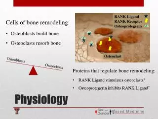
MEMBRANE PROTEINS • Each cell: 10-50 different types of membrane proteins. • Proteins determine membrane’s specific functions. • Cell membrane & organelle membranes each have unique collections • of proteins. • Peripheral proteins (Cell surface identity marker (antigens)) • present either to the outside or inside of the cell. • Some of them are anchored to the membrane by covalent bonding. • Others held non-covalently that can be disrupted by gentle movement or change in pH. • Include enzymes and binding proteins that anchor cell to membrane. • Integral proteins (Transmembrane proteins) • Tightly bound to phospholipid bilayer. • Present within the matrix of the membrane.
oligosaccharide groups cholesterol phospholipid EXTRACELLULAR ENVIRONMENT (cytoskeletal pro-teins beneatch the plasma membrane) open channel protein gated channel proten (open) gated channel proten (closed) active transport protein RECEPTOR PROTEIN LIPID BILAYER ADHESION PROTEIN RECOGNITION PROTEIN (area of enlargment) TRANSPORT PROTEINS CYTOPLASM PLASMA MEMBRANE
Membrane carbohydrates • Play a key role in cell-cell recognition (The carbohydrate chains of glycolipids and glycoproteins serve as the “fingerprints” of the cell). • Glycolipids and glycoproteins vary from species to species and even from cell to cell in the same individual. • ability of a cell to distinguish one cell from another. • important in organ & tissue development. • basis for rejection of foreign cells by immune system. • Person’ particular blood group is due to the presence of particular glycoproteins in the membrane of RBC.
Fluidity of plasma membrane • At body temperature plasma membrane has the consistency of olive oil. • The greater the concentration of unsaturated fatty acids residues, the more fliud is the bilayer. • The hydrocarbon tail of phospholipid molecule wiggle and the entire phospholipid molecule can move sideways at a rate of 2µm per second. • Some phospholipids rarely flip-flop from one layer to the other. • Cholesterol which iswedged between phospholipids molecules in the plasma membrane of animals cells, restrains the movement of the phospholipids in warm temps and maintains fluidity by preventing tight packing at cold temps thereby add support and rigidity to the membrane.
functions • Physical isolation - Separates inner and outer environment, surrounds & gives cell a particular shape. • Lipids restricts passage of polar compounds. • Proteins customize membranes • Provide structural support • Serve as transporters, enzymes, receptors & identity markers • Regulates exchange with the environment - membrane channel proteins + carrier proteins. • Carbohydrates in form of glycoproteins & glycolipids are part of outer surface Impart negative charge to surface (cell recognition) (glycocalyx). • Allows a steady supply of glucose, amino acids, and lipids to come into the cell. • Maintains concentration gradient across the membrane. • Maintains electrochemical gradient across the membrane. • “homeostasis”Sensory receptor - membrane receptor proteins sense changes in external environment (encrusted with peripheral nerves).
Other components of the plasma membrane Cholesterol plays the important role of preventing the fatty acid chains of the phospholipids from sticking together. Cholesterol Molecule
Protein Functions • Channel Proteins - Involved in passage of molecules or ions through membrane. • Carrier Proteins - Combine with substance to aid in passage through membrane. • Cell Recognition Proteins - Help body recognize foreign substances. MHC (major histocompatabilty complex) glycoproteins, are difficult for each person, therefore rejected at the time of organ transplantation. • Receptor Proteins – Shaped in such a way that a specific molecule can bind to it. It may sometimes cause protein to change shape and bring about the transport. • Enzymatic Proteins – Catalyse a specific reaction.(adenylate cyclase involved in ATP synthesis)
Classes of Amino acids What do these amino acids have in common? Nonpolar & hydrophobic
Classes of amino acids What do these amino acids have in common? Polar & hydrophilic
Proteins domains anchor molecule Polar areas of protein • Within membrane • nonpolar amino acids • hydrophobic • anchors protein into membrane • On outer surfaces of membrane • polar amino acids • hydrophilic • extend into extracellular fluid & into cytosol Nonpolar areas of protein
Transport Across Plasma Membrane • Selectively permeable. • Passive • Diffusion, • Osmosis, • Facilitated diffusion • Active • Primary active transport, • Secondary active transport.
Diffusion • Movement of molecules from higher concentration to a lower concentration( down the gradient), until equilibrium is attained. • Non-polar highly polar compounds readily diffuse through cell membrane. • Most drugs follow this type of transport.(90%) • Size of molecule 100-200 D. • First order kinetics. • ex: Fatty acids, steroids vitamins, etc.,
Osmosis (pore transport, aquaporins) • The process of water moving from low solute concentration to high solute concentration across a semi-permeable membrane is called osmosis. • Upto 100 D, 0.4nm. • Ex: Highly water soluble drugs, drugs entering liver, renal excretion, removal of drugs from CSF. Solute: particles dissolved in water : Fe, Mg, O2, etc.
Ion channels • Charged & most polar compounds must have an ion channel or transporter to move across membrane • Ion channels are integral proteins, which conduct ions like Na+, K+ ,Ca+2 • They can differentiate between the size and charge of the ion that is being diffused. • They may open or gated and depend on chemical, electrical or mechanical signals for the transfer of a specific molecule across the membrane.
Facilitated diffusion • Involves special carrier protein for the transfer of molecule across the membrane. • These carriers are specific only to a particular molecule. • Once the molecule binds to the protein a conformational change occurs in the protein, which moves the molecule down its concentration gradient. • Saturation or transport maximum. • Specific • Competition • Mixed order kinetics.
Active transport • Across the concentration gradient. • Consumes 40% of the cell energy. • Selective • Saturation • Competition. • Specific in location. • Ex: thiamine, nicotinic acid, riboflavin, pyridoxine, 5-floro uracil, methyl dopa, nicotinamide. • Two types: Primary Secondary
Primary Active Transport: 1) Na+/K+ Pump: • Present in thousands per cell. • Helps maintain sodium on the outside and potassium inside. • When three Na+ ions bind to the protein the ATP-pase function of the protein becomes activated. splitting of ATP to ADP and high energy phosphate. High energy phosphate causes conformational change helps moving Na+ ions outside of the cell and brings 2 K + ions into the cell.
Secondary Active Transport: • Co-transport or Symport: The diffusion gradient of Na+can pull other molecules along with it, this is called co-transport or symport. ex: glucose-sodium pump, lactose-proton pump, sodium-amino acid pump, sodium-proton pump. • Antiport or Counter transport: The diffusion gradient of Na+ exchanges other molecules with it, this is called Antiport or Counter transport. 1) Na+/ Ca+ pump
Transport of large particles Endocytosis- A cell surrounds material and takes it in from its environment by enclosing it in a newly formed vacuole. 3 types: 1) Receptor-mediated endocytosis-vitamins antibodies,hormones, virus. 2) Phagocytosis. 3) Bulk phase endocytosis.
Transport of drugs • 2 super families. • ABC (ATP binding cassette)-active • SLC (solute carrier)-facilitated ABC: • Active transport • Divided into 7 subclasses (or) families (A-G) These include 1) P-gp (encoded as ABCB1/MDRI). 2) Cystic Transmembrane CFTR regulator(ABCC7). SLC: • Facilitated Transport. • Ion coupled. • 43 SLC Families, 300 Transporters. • Seratonin SERT, (SLC6A4), Dopamine Transporters, DAT(SLC6A3).
Factors responsible for drug absorption across the membrane • Molecular size • Shape • Degree of ionization • Surface area of exposed membrane exposed. • Relative solubility of ionized & unionized forms. • Concentration gradient across the membrane. • Receptor availabilty.
References • Principles of Anatomy & Physiology, Gerard.J.Tortora, Bryan Derrickson. • Pharmacology, H.P.Rang, M.M.Dale. • The Pharmacological Basis Of Therapeuctics, Goodman’s and Gilman’s. • Biopharmaceutics and Pharmacokinetics, D.M.Bramhankar, Sunil B.Jaiswal. • www.sciencedirect.com • www.pubmed.com • www.pharmacorama.com • THANK YOU