
Hypersensitivity Reactions: Mechanisms, Diagnosis, Treatment
E N D
Presentation Transcript
HYPERSENSITIVITY REACTIONS DR.JOEL SOLORZANO ROMERO
TEACHING OBJECTIVES: 1. Understand the classification of hypersensitivity reactions 2. Know the diseases associated with hypersensitivity reactions 3. Understand the mechanisms of damage in hypersensitivity reactions 4. Know the methods for diagnosing conditions due to hypersensitivity 5. Know the modes of treating disease due to hypersensitivity and their rationale
INTRODUCTION Origins of Hypersensitivity “Hypersensitivity” first used clinically in 1893: • attempting to protect against diphtheria toxin • test animals suffered enhanced responses,even death following second toxin exposure • at miniscule doses not harmful to untreated animals The term “Allergy” is coined in 1906: postulated to be the product of an “allergic” response • from Greek allosergos (altered reactivity)
Hypersensitivity: Aberrant or excessive immune response to foreign antigens • Primary mediator is the adaptive immune system T & B lymphocytes • Damage is mediated by the same attack mechanisms that mediate normal immune responses to pathogen
Common to All Types Adaptive (T & B Cell) Immune Responses • Reactions occur only in sensitized individuals • Generally at least one prior contact with the offending agent • Sensitization can be long lived in the absence of re-exposure (>10 years) due to immunologic memory • Antigen is a protein or is capable of complexing with protein (hapten)
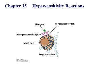
TYPE 1 HYPERSENSITIVITY Type 1 hypersensitivity reaction is an allergic reaction provoked by re-exposure to a specific type of antigen referred to as an allergen. The reaction may be either local or systemic. Symptoms vary from mild irritation to sudden death from anaphylactic shock. Exposure may be by ingestion, inhalation, injection, or direct contact. The ‘naïve’ b lymphocytes become primed and differentiate into an antibody–secreting cell and this leads to class-switching of the antibody to the IgE class
The difference between a normal immune response and a type I hypersensitivity response is that plasma cells secrete IgE. This class of antibodies binds to Fc receptors on the surface of tissue mast cells and blood basophils. Mast cells and basophils coated by IgE are “sensitised” Later exposure to the same allergen, cross-links the bound IgE on sensitised cells resulting in degranulation and the secretion of pharmacologically active mediators such as histamine, leukotriene,and prostaglandins that act on the surrounding tissues. The principal effects of these products are vasodilation and smooth-muscle contraction
Sensitization • Antigen contact, typically low-dose via mucous membranes (respiratory, GI) IgE production Elicitation (Re-exposure • Pre-formed IgE (allergen-specific) triggers mast cell activation ⌫ mediator release Reactions • Can occur within seconds-minutes of exposure • Severity ranges from irritating to fatal IgE Production
IgE Production Secondary immune response (multiple or persistent exposures) B cell class switch to IgE requires T cell help:CD40L and IL-4 or IL-13 (Th2 cytokines) The propensity to make an IgE response to environmental antigens varies among individuals ”Atopic” individuals are those with an inherited predisposition to form IgE responses
Sensitization Response IgE produced by plasma cells is rapidly taken up by Fc on tissue mast cells and circulating basophils (serum τ½~2 days; compare to IgG~21 days) Immediate Histamine (also tryptase, heparin) Smooth muscle constriction Vasodilatation; vascular leak G.I. motility (increased) Mucous Secretion Sensory nerve activation Early Phase: IgE crosslinking by antigen release of preformed mediators
Early Phase: Followed by rapid production of arachidonic acid products. Minutes Leukotrienes, prostaglandins Smooth muscle constriction Vasodilatation; vascular leak Mucous Secretion Neutrophil chemotaxis
Late Phase: Gene activation new cytokine production ~6 hours after antigen triggering Cytokines TNFα recruit inflammatory cells IL-3, IL-5, GM-CSF eosinophil production IL-4, IL-13 propogate Th2 respo
Anaphylaxis Response to systemic circulation of allergen • IgE cross-linking on mast cells in peri-vascular tissue • Circulating histamine, PG’s/LT’s ➟ vasodilatation, vascular leak • High-output shock: ↓↓BP despite ↑’ed cardiac output • Other symptoms: urticaria, wheeze, laryngeal edema with airway compromise, G.I. cramping, diarrhea, “feeling of dread” • Symptoms progress rapidly (seconds) • Treatment • Immediate: epinephrine 0.3 cc s.c, followed by antihistamines (H1 and H2 blockade) IM or IV ➟
Some examples of type 1 hypersensitivity: Allergic asthma Allergic conjunctivitis Allergic rhinitis (“hay fever”) Anaphylaxis Angioedema Atopic dermatitis (eczema) Urticaria (hives) Eosinophilia
Symptomatic treatment is achieved with antihistamines which block histamine receptors. Chromolyn sodium inhibits mast cell degranulation, probably, by inhibiting Ca++ influx. Late onset allergic symptoms, particularly bronchoconstriction which is mediated by leukotrienes are treated with leukotriene receptor blockers (Singulair,Accolate) or inhibitors of cyclooxygenase pathway . Symptomatic, although short term relief from bronchoconstriction is provided by bronchodilators (inhalants) such as isoproterenol derivatives (Terbutaline,Albuterol). Thophylline elevates cAMP by inhibiting cAMP-phosphodiesterase and inhibits intracellular Ca++ release is also used to relieve bronchopulmonary symptoms.
TYPE 2 HYPERSENSITIVITY In type 2 hypersensitivity reactions, the antibodies produced by the immune response bind to antigens on the patient’s own cell surfaces. The antigens recognised in this way may either be intrinsic (“self” antigen, innately part of the patient’s cells) or extrinsic (absorbed onto the cells during exposure to some foreign antigen, possibly as part of infection with a pathogen).
IgG and IgM antibodies bind to these antigens to form complexes that activate the classical pathway of complement activation, for eliminating cells presenting foreign antigens (which are usually, but not in this case, pathogens). That is, mediators of acute inflammation are generated at the site and membrane attack complexes cause cell lysis and death. The reaction takes hours to a day
Phagocytosis of the cell can be mediated by phagocytes expressing Fc receptors. These Fc receptors recognise surface bound antibody and complement receptors, that recognise surface bound complement protein. These cells are also recognised by macrophages or dendritic cells which act as antigen presenting cells, this causes a B cell response where antibodies are produced against the foreign antigen.
Another form of type 2 hypersensitivity is called Antibody Dependent Cell Mediated Cytotoxicity (ADCC). Here, cells exhibiting the foreign antigen are tagged with antibodies (IgG or IgM). These tagged cells are then recognised by Natural Killer (NK) cells and macrophages (recognised via IgG bound to the cell surface receptor, CD16 (Fc?RIII)), which in turn kill these tagged cells
Some examples: Autoimmune hemolytic anemia Goodpasture’s syndrome Erythroblastosis Fetalis Pemphigus Pernicious anemia (if autoimmune) Immune thrombocytopenia Transfusion reactions Hashimoto’s thyroiditis Graves’ disease (see type V below) Myasthenia gravis (see type V below) Rheumatic fever Hemolytic disease of the newborn
Type 3 – Immune Complex In type 3 hypersensitivity reactions, soluble immune complexes (aggregations of antigens and IgG and IgM antibodies) form in the blood and are deposited in various tissues (typically the skin, kidney and joints) This deposition of the antibodies may trigger an immune response according to the classical pathway of complement activation – for eliminating cells presenting foreign antigens (which are usually, but not in this case, pathogens). There are two stages relating to the development of the complexes, firstly the complex forms when IgG and IgM antibodies are bound to an antigen, after this, the complexes can form larger ones which can be cleared by the body
It is also known as immune complex hypersensitivity. The reaction may be general (e.g., serum sickness) or may involve individual organs including skin (e.g., systemic lupus erythematosus, Arthus reaction), kidneys (e.g., lupus nephritis), lungs (e.g., aspergillosis), blood vessels (e.g., polyarteritis), joints (e.g., rheumatoid arthritis) or other organs. This reaction may be the pathogenic mechanism of diseases caused by many microorganisms.
Tissue damage results at the site of the immune complex with the influx of phagocytes and granuloctyes and the release of inflammatory mediators Some examples: Immune complex glomerulonephritis Rheumatoid arthritis Serum sickness Subacute bacterial endocarditis Symptoms of malaria Systemic lupus erythematosus Arthus reaction Farmer’s Lung (Arthus-type reaction)
Serum Sickness 2-3 weeks following infusion of antigen (classically an antiserum of horse origin) Fever Lymphadenopathy Urticaria Joint Pain Proteinuria
Arthus reaction In immunology, the Arthus reaction is a type of local type III hypersensitivity reaction. Type III hypersensitivity reactions are immune complex mediated, and involve the deposition of an antigen/antibody complex mainly in the vascular walls, serosa (pleura, pericardium, synovium), and glomeruli The Arthus reaction involves the in situ formation of antigen/antibody complexes after the intradermal injection of an antigen. If the animal/patient was previously sensitised (has circulating antibody), an Arthus reaction occurs. This manifests as local vasculitis due to deposition of immune complexes in dermal blood vessels. Activation of complement and recruitment of PMNs ensue which results in an inflammatory response.
Arthus reactions are characterised by severe pain, swelling, induration, edema, hemorrhage, and occasionally by necrosis. These symptoms and signs usually occur 4–12 hours after vaccination.
Type 4 – Cell-mediated (Delayed-Type Hypersensitivity, DTH) Type 4 hypersensitivity reactions are often called delayed type as the reaction takes two to three days to develop. Unlike the other types, it is not antibody mediated but rather is a type of cell mediated response. CD8+ cytotoxic T cells and CD4+ helper T cells recognise antigen in a complex with either type 1 or type 2 major histocompatibility complex. The antigen-presenting cells in this case are macrophages which secrete IL-1, which stimulates the proliferation of further CD4+ T cells
Re-exposure to the allergen results in a Th1 mediated response which stimulates the proliferation of the allergen-specific memory Th1 CD4+ T helper lymphocyte via recognition of complexes of peptide on antigen presenting cells (APCs) CD4+ T cells secrete IL-2 and interferon gamma, further inducing the release of other Type 1 cytokines, thus mediating the immune response. Activated CD8+ T cells destroy target cells on contact while activated macrophages produce hydrolytic enzymes and, on presentation with certain intracellular pathogens, transform into multinucleated giant cells
Diseases associated with granuloma formation: Leprosy Tuberculosis Schistosomiasis Sarcoidosis Crohn’s disease
Some examples: Contact dermatitis (poison ivy rash, for example) Temporal arteritis Symptoms of leprosy Symptoms of tuberculosis Transplant rejection Coeliac disease