Download
1 / 29
290 likes | 352 Vues
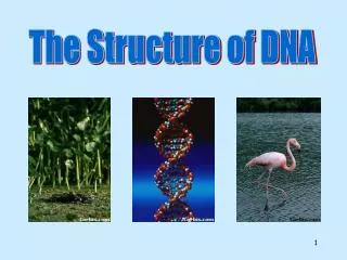
Explore the intricacies of DNA, the double helix structure, nucleic acid composition, nucleotide components, nitrogenous bases, and historical breakthroughs in genetics. Understand DNA packaging, karyotypes, and pivotal experiments that unveiled DNA's role as the genetic material.

E N D
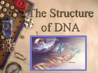
Deoxyribonucleic acid (DNA) Nucleic Acid – Polymer monomer = nucleotide. 2 kinds of nucleic acids: DNA and RNA.
Coding sections in DNA are called genes. • Genes code for protein production; • (the hereditary info. in DNA tells cells how to make proteins)
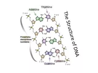
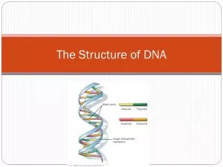
Nucleotide: three components (CP: 230) (H: 197) 5 C sugar (deoxyribose) phosphate group nitrogenous base(4 kinds in DNA) Thymine Adenine Guanine Cytosine
Nucleotide: three components (CP: 230) (Hon: 197) 5 C sugar phosphate group nitrogenous base(4 kinds in DNA)
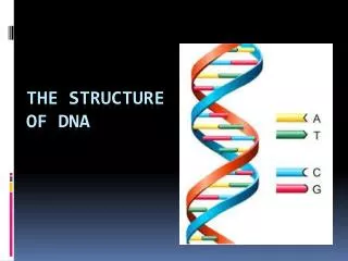
O N N O N N N N C C C C N O C C C C C C C C O C N N N N C C N N C C Cytosine C Guanine Thymine Adenine C N N Four Kinds of Nitrogenous Bases • Adenine binds to Thymine • Cytosine binds to Guanine • A-T and C-G (always)
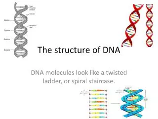
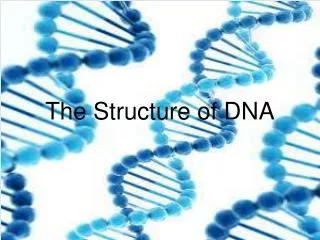
DNA STRUCTURE Shape is like twisted ladder. Called a double helix -two twisted strands Bases =rungs of a ladder hydrogen bonds hold pairs together; A-T, C-G Sugars- phosphates form sides of ladder. Covalent bonds in backbone
Carbons in the sugar are labeled 1’ (one prime) to 5’.
Model parts: • 12 – White Tees [Deoxyribose] • 6 – White connectors [Hydrogen bond] • 12 – Black [Phosphate group] • 3 – Green [Guanine] • 3 – Blue [Adenine] • 3 – Red [Thymine] • 3 – Yellow [Cytosine]
DNA Animation http://www.johnkyrk.com/DNAanatomy.html
Packaging DNA Fundamental unit is Nucleosome – • DNA wound around proteins called histones. • (Hon. page 151) • Occurs at Prophase
Nucleosomes • Nucleosomes • Lowest DNA packaging level • thread wound around a spool
DNA - By The Numbers! • Each cell has about 2 meters (6 ft) of DNA. • The average human has 60-75 trillion cells. • Avg human has enough DNA to go from the Earth to the sun more than 400 times. • DNA has a diameter of only 0.0000000002 meters (20Ǻ) [1Ǻ =10-10 m] The earth is 150 billion meters or 93 million miles from the sun.
Karyotypes • Normal human male karyotype(the total set of chrom. of an organism)
Karyotypes • Normal human female karyotype
The History of DNA • Gregor Mendel-1866 • Determined “Unit characters” were the method of passing on traits for inheritance • Friedrich Meischer - 1868 • Studied nuclei of pus cells obtained from discarded surgical bandages • Detected a phosphorus-containing substance that he named nuclein.
Frederick Griffith 1928 : Work with Bacteria Found that DNA taken from a virulent (disease-causing) strain of bacteria (Streptococcus pneumoniae) Transformed a non-virulent form of the bacterium into a virulent form.
Oswald Avery, Colin MacLeod, and Maclyn McCarty 1943Continued the study of “Transformation” principle
Meischer Mendel Griffith MacLeod McCarty Avery
Transformation Of BacteriaTwo Strains Of Streptococcus Capsule Rough Strain (Harmless) Smooth Strain w/Capsule (Virulent)
Control Control Control Experimental Transformation Of Bacteria -Griffith’s Experiment Smooth (virulent) OUCH! Rough
The History of DNA • Alfred Hershey and Martha Chase - 1952 • radioactive isotope tracer experiment • bacterial virus (bacteriophageT2) infects a host cell ( bacterium Escherichia coli) • found that T2 virus DNA, not its protein coat, enters the host cell • genetic information for replication of the virus
T 2 grown in media containing S35 incorporate S35 into their proteins Bacteria grown in normal non-radioactive media T2 attach to bacteria and inject genetic material Mix-O-Matic Blending causes phage protein coat to fall off Using S35 When centrifuged, phage protein coats remain in the supernatant while bacteria form a pellet. The supernatant is radioactive, but the pellet is not. Did protein enter the bacteria? Is protein the genetic material?
T2 grown in P32 containing media incorporate P32 into their DNA Bacteria grown in normal non-radioactive media T2 attach to bacteria and inject genetic material Mix-O-Matic Blending causes phage protein coat to fall off Using P32 When centrifuged, phage protein coats remain in the supernatant while bacteria form a pellet The pellet is radioactive, but the supernatant is not. Did DNA enter the bacteria? Is DNA the genetic material?
The History of DNA Structure • Erwin Chargaff- 1940 - “Chargaff’s rule” • four bases may occur in varying proportions in DNA of different organisms • # of A = # of T, w/ two hydrogen bonds • = # of G and C are present w/ 3 hydrogen bonds • Rosalind Franklin and Maurice Wilkins • X-ray diffraction study concluded DNA fibers have two strands.
The History of DNA Structure • James Watson and Francis Crick - University of Cambridge -1953 • Worked on problem of making a DNA molecule model that was double stranded but also had the specific A - T and G - C base equivalencies • Solution-double helical structure for DNA.
Franklin Chargaff Wilkins