Download

1 / 26
350 likes | 1.67k Vues
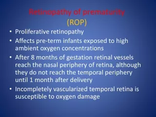
Retinopathy of Prematurity. Learning Activity 2 By: Scarlett Bayard Kandice Burke Summer Gibson. What is ROP?.

E N D
Retinopathy of Prematurity Learning Activity 2 By: Scarlett Bayard Kandice Burke Summer Gibson
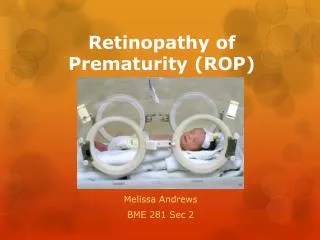
What is ROP? Retinopathy of prematurity (ROP) is a disease that primarily occurs in premature babies. It causes abnormal blood vessels to grow in the retina, the layer of nerve tissue in the eye that enables us to see. This growth can cause the retina to detach from the back of the eye, leading to blindness.
What is ROP continued… Retinopathy of Prematurity (ROP), originally called retrolental fibroplasia, was the leading cause of blindness in children in the 1940s and 1950s. ROP occurs when abnormal blood vessels grow and spread throughout the retina, the tissue that lines the back of the eye. These abnormal blood vessels are fragile and can leak, scarring the retina and pulling it out of position. This causes a retinal detachment. Retinal detachment is the main cause of visual impairment and blindness in ROP.
Interesting Facts Retinopathy of prematurity (ROP) is a potentially blinding eye disorder that primarily affects premature infants weighing about 2¾ pounds (1250 grams) or less that are born before 31 weeks of gestation (A full-term pregnancy has a gestation of 38–42 weeks). The smaller a baby is at birth, the more likely that baby is to develop ROP. This disorder—which usually develops in both eyes—is one of the most common causes of visual loss in childhood and can lead to lifelong vision impairment and blindness. ROP was first diagnosed in 1942. ROP occurs in over 16% of all premature births. In babies weighing less than 1,700 grams at birth, over 50% will develop ROP. In the United States, over 2,100 children annually experience the complications of ROP. Of those estimates of 500 to 1,200 cases of new blindness or severe complications are reported. Studies have found that about 30% of infants with advanced ROP have 20/200 or less in their better eye.
How is the Visual System Affected? Several complex factors may be responsible for the development of ROP. The eye starts to develop at about 16 weeks of pregnancy, when the blood vessels of the retina begin to form at the optic nerve in the back of the eye. The blood vessels grow gradually toward the edges of the developing retina, supplying oxygen and nutrients. During the last 12 weeks of a pregnancy, the eye develops rapidly. When a baby is born full-term, the retinal blood vessel growth is mostly complete (The retina usually finishes growing a few weeks to a month after birth). But if a baby is born prematurely, before these blood vessels have reached the edges of the retina, normal vessel growth may stop. The edges of the retina—the periphery—may not get enough oxygen and nutrients.
Visual System Affected continued… Scientists believe that the periphery of the retina then sends out signals to other areas of the retina for nourishment. As a result, new abnormal vessels begin to grow. These new blood vessels are fragile and weak and can bleed, leading to retinal scarring. When these scars shrink, they pull on the retina, causing it to detach from the back of the eye. Today, we realize that oxygen is not the only factor in developing ROP. High levels of oxygen have been associated with ROP, but lower levels of oxygen may lead to more respiratory complications and death in premature infants. Better oxygen level monitoring has led to better control of the oxygen given to premature infants. Today, however, there is an increase in ROP due to the fact that neonatal care advances mean more low weight premature infants are surviving.
Effects on the Visual System Retinal detachment (RD) is common in patients with ROP. In many cases, it leads to profound vision loss. The retina is the light sensitivity “film-like” portion of the eye. A retinal detachment occurs when this delicate tissue is dislodged from the internal walls of the eye. Early detection and treatment is crucial. In stage 5, the most severe form of ROP, retinal detachment surgery may not be attempted due to the poor prognosis versus risk of operating on a premature infant.
Visual System Effects continued… Strabismus is another complication and it is the crossing in or turning out of an eye. This may occur from the loss of vision in one eye or be related to the large refractive differences between the eyes. This difference is called an anisometropia or antimetropia. For example, one eye may be 1 unit of myopia (nearsightedness) while the other eye is six units of nearsightedness. Both cataract and corneal problems can develop. Severe damage may lead to Phthisis bulbi, a shrinking of the severely damaged eyes. Glaucoma may develop either early or as a later in life complication of ROP. Additionally, ROP patients may have high amounts of nearsightedness called myopia. In patients who experience significant vision loss, nystagmus, a rapid fluttering of the eyes, may occur. Nystagmus is common in all patients whose vision loss occurs at an early age.
ROP Classification ROP is classified in five stages, ranging from mild (stage I) to severe (stage V): • Stage I — Mildly abnormal blood vessel growth. Many children who develop stage I improve with no treatment and eventually develop normal vision. The disease resolves on its own without further progression. • Stage II — Moderately abnormal blood vessel growth. Many children who develop stage II improve with no treatment and eventually develop normal vision. The disease resolves on its own without further progression. • Most babies who develop ROP have stages I or II. However, in a small number of babies, ROP worsens, sometimes very rapidly. Untreated ROP threatens to destroy vision.
ROP Classification continued… Stage III — Severely abnormal blood vessel growth. The abnormal blood vessels grow toward the center of the eye instead of following their normal growth pattern along the surface of the retina. Some infants who develop stage III improve with no treatment and eventually develop normal vision. However, when infants have a certain degree of Stage III and "plus disease" develops, treatment is considered. "Plus disease" means that the blood vessels of the retina have become enlarged and twisted, indicating a worsening of the disease. Treatment at this point has a good chance of preventing retinal detachment. Stage IV — Partially detached retina. Traction from the scar produced by bleeding, abnormal vessels pulls the retina away from the wall of the eye.
ROP Classification continued… Stage V — Completely detached retina and the end stage of the disease. If the eye is left alone at this stage, the baby can have severe visual impairment and even blindness.
Treatments and Medications ROP surgery is used to stop the growth of abnormal blood vessels by focusing treatment on the peripheral retina (the sides of the retina) to preserve the central retina (the most important part of the retina). ROP surgery involves scarring areas on the peripheral retina to stop the abnormal growth and eliminate pulling on the retina. Since surgery focuses treatment on the peripheral retina, these areas will be scarred and some amount of peripheral vision may be lost. However, by preserving the central retina, the eye will still be able to perform vital functions like seeing straight ahead, distinguishing colors, reading, etc.
Treatments & Medications continued… • Both laser treatments and cryotherapy are performed only on infants with advanced ROP, particularly stage III with "plus disease." Both treatments are considered invasive surgeries on the eye, and doctors don't know the long-term side effects of each. In the later stages of ROP, other treatment options include: • Scleral buckle:This involves placing a silicone band around the eye and tightening it. This keeps the vitreous gel from pulling on the scar tissue and allows the retina to flatten back down onto the wall of the eye. Infants who have had a sclera buckle need to have the band removed months or years later, since the eye continues to grow; otherwise they will become nearsighted. Sclera buckles are usually performed on infants with stage IV or V. • Vitrectomy:Vitrectomy involves removing the vitreous and replacing it with a saline solution. After the vitreous has been removed, the scar tissue on the retina can be peeled back or cut away, allowing the retina to relax and lay back down against the eye wall. Vitrectomy is performed only at stage V.
Congenital or Adventitious? Retinopathy of Prematurity is a congenital disorder. If an infant is born premature, it does not mean they will acquire ROP. However, ROP is more likely to occur with very small and sick infants. ROP is rarely seen in infants that weigh more than 2.75 lbs. at birth.
Progressive or Stable? ROP is usually mild, and typically it will disappear on its own weeks after it developed. About 98% of babies screened for ROP will not need treatment. ROP that is mild typically goes away with no outside help. If treatment is needed, around 90% will have the ROP vanish, and their vision will be fine.
Anticipated Functional Implications What can you do in their daily life for them? Use a multisensory teaching approach that could combine use of vision with auditory, tactual, and kinesthetic input. Use real objects and engage the child in daily activities that occur in life. Describe pictures, actions, body language, gestures, and sounds as best as possible. Assist them in developing good listening skills.
More Functional Implications What could help your child at school? • Use a slant board • Light box • Help the student learn to scan their environment • High contrast materials • Evaluate the child’s abilities as they grow to see if the ROP has changed • Braille should be considered • Low Vision Evaluation • Preferential seating – exact location to be specified
Case Study: Sam Etiology: Retinopathy of Prematurity Age: 4 yrs old Onset: congenital
Sam’s Parent Interview History through Parent Interview: Sam’s mother had a normal pregnancy. At 21 weeks of pregnancy she received steroid shots to strengthen the twins’ lungs. Three weeks later (24 weeks) she was admitted to the hospital. Sam was born at 25 weeks weighing 1lb, 9oz. Soon after birth Sam developed NEC which is, Necrotizing Entercolitis. NEC affects 2% of premature babies. It is an infection of the intestines which causes inflammation, interior abdominal damage, and tissue death. Sam received several surgeries for the NEC. At 36 weeks gestation Sam’s parents were told that he had Retinopathy of Prematurity (ROP.) Between 36 and 40 weeks gestation Sam received two surgeries in his right eye and one in his left eye. Doctors cauterized the blood vessels so that they would not detach the retina. Sam came home from the hospital 9 months after he was born. He came home on oxygen, with a trach, feeding tube/pump, and nursing care 80 hrs per week. When asked how she knew what “to do next” Sam’s mother stated that the NICU nurse talked them through what to do (ECI.) They also attended parent/family support group meetings through the hospital every 2 weeks.
Sam’s Medical History Medical History Report states: Sam’s medical history includes chronic lung disease, which required that he receive around the clock oxygen until he was approximately 18 months old, Retinopathy of Prematurity, NEC (feeding tube), PDA ligation, and 2 level 2 brain bleeds. Sam’s tracheotomy and feeding tube have both been removed. Sam received laser surgery on both eyes for the ROP and the doctor reported that the surgery had a good result. Dr. Stager Jr. suggested surgery for Nystagmus however; mom states that it has dampened. Dr. Stager Jr. also suggested patching the left eye in order to strengthen the right eye. This is done for 30 minutes daily. Sam’s medical diagnosis is H/O ROP, Myopic Astigmatism, Nystagmus. There is no apparent field restriction and Sam is not legally blind. Sam does not have serious visual loss after correction. Eye glasses were recommended.
Sam’s Evaluation and Assessment Functional Vision Re-Evaluation and Learning Media Assessment: Sam was tested in various rooms within his home, hallway, outside the school, occupied office, unoccupied classroom and the play-based assessment room at Acker Special Programs Center. During the evaluation Sam was able to easily track a toy and a penlight horizontally at 12 inches. He was also able to track vertically at 12 inches. Sam was able to track a 6 inch blue ball rolling across the floor at a distance of 3-5 feet. Sam responded to all objects in his peripheral fields and in his upper and lower quadrants. Although Sam is a non-reader due to his age he is showing some prerequisite signs of reading. His writing skills are emerging. Sam was able to scan the room to find a large 10 inch blue ball at 15 feet when asked to do so. He tracked a smaller 6 inch ball at 5 feet. He saw his mom at a distance of 10-15 feet but it was undetermined whether or not he recognized her based on visual cues only until he was approximately 5 feet from her. It was observed, though inconsistently, that at time Sam demonstrates some eccentric viewing when looking at object/people at a distance. He tilts his head up and to the side and looks out of the lower corners of his eyes. This may be Sam’s attempt to find the null point due to his Nystagmus. Based on the Functional Vision Evaluation, student behaviors, teacher interview, parent interview, and the eye doctor’s report, Sam’s primary sensory channel for learning is visual with auditory as the secondary learning mode. Sam is not functionally blind and does not require braille. His most appropriate reading medium is print. For writing his most appropriate medium is pencil and paper. It is undetermined at this time if low vision devices will be needed.
Sam’s Educational Implications Educational Implications: Sam meets the criteria as visually impaired in accordance with the Texas Education Agency (TEA) guidelines. Based on visual performance and review of eye medical reports, even with correction, Sam has an impairment in vision that adversely affects child’s educational performance creating a need for special education (i.e. specially designed instruction). It is recommended that Sam receive vision services in order to implement visual modifications and instructional approaches. Several adaptions and/or modifications are appropriate for Sam. Sam will appear to be looking in different direction to accommodate his peripheral viewing. Preferential seating may be necessary. Encourage Sam to move within classroom to accommodate viewing needs. Present visual material in a simple uncluttered format. A multi-sensory approach to learning should be used whenever possible. For near visual tasks Sam should use real objects whenever possible. For reading activities, Sam needs: high contrast print with maximum spacing should be used when possible, large font on computer is recommended. For writing activities, Sam may need to utilize bold line paper and may require a felt tip pen or a dark leaded pencil. For math activities, Same may need to use actual objects (i.e. Cuisenaire rods) for teaching counting or introducing new concepts, rather than pictures and use real money to teach money concepts. Sam may have problems with glare (i.e. reduce glare on chalkboard/TV.) He may need sunglasses and/or visors when outdoors/indoors. Sam has difficulty adjusting to changes in light, will need time to adapt (i.e. inside to outside.)Teacher should not stand with back to window. Sam should not look directly into window. Reduce glare on chalkboard/TV. No assistive technology is recommended at this time.
Sam’s Services Services: VI services: 1xmonthly Orientation & Mobility services: 1xmonthly Speech services: 1hr/month Attends the Early Childhood School: 5xweekly for ½ day Special Education transportation
References • Retinopathy of prematurity. (2010, July 05). Retrieved from http://www.lowvision.org/retinopathy_of_prematurityxx.htm • About retinopathy of prematurity. (2010, July 05). Retrieved from http://kidshealth.org/parent/system/surgery/rop.html • Facts about retinopathy of prematurity. (2010, July 05). Retrieved from www.nei.nih.gov/health/rop.asp • Educating students with visual impairments for inclusion in society. (2010, July 05). Retrieved from http://www.afb.org/Section.asp?SectionID=44&TopicID=189&DocumentID=1344 • Keller, Ed. (2005). Strategies for teaching visually impaired students. Retrieved from http://www.as.wvu.edu/%7Escidis/vision.html#sect3 • Lueck, Amanda. (2004). Functional vision. Amer Foundation for the Blind. • Lindquist, Sonja (n.d.). Necrotizing Enterocolitis (NEC) and Your Premature Baby. Retrieved July 21, 2010, from http://ezinearticles.com/?Necrotizing-Enterocolitis-(NEC)-and-Your-Premature-Baby&id=309221.