Download

1 / 30
490 likes | 1.77k Vues
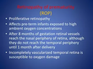
Retinopathy of Prematurity. Meena Kumar 10/1/03. Definition. Retinopathy of prematurity (ROP) was formerly known as retrolental fibroplasia. It is a developmental vascular proliferative disorder that occurs in the incompletely vascularized retina of primarily premature infants.

E N D
Retinopathy of Prematurity Meena Kumar 10/1/03
Definition • Retinopathy of prematurity (ROP) was formerly known as retrolental fibroplasia. • It is a developmental vascular proliferative disorder that occurs in the incompletely vascularized retina of primarily premature infants. • ROP is one of the most common causes of blindness in children.
Incidence of ROP • Hussain et al. (Pediatrics 1999; 104:e26): 950 infants born between 1989 and 1997 had serial eye exams if they were less than 30 wks GA, less than 1300g BW, or required O2 supplementation. • Incidence of ROP increased with decreasing GA or decreasing BW.
Vascular Development of the Eye Nasal side Temporal side
Pathogenesis • Not well understood but thought to involve two stages: • Stage I: an initial injury (such as hypotension, hypoxia, or hyperoxia) causes vasoconstriction and reduced blood flow to the retina, disrupting the normal process of vascularization. • Stage II: Vessels then either resume normal growth or new vessels grow abnormally out from the retina into the vitreous. The abnormal vessels have increased permeability which can result in edema and hemorrhage. Inflammation → fibrous tissue → traction on the retina and detachment. Alternatively, the abnormal vascularization may regress with little residual effect. • Recently, the interaction between insulin-like growth factor-1 (IGF-1) and VEGF has been studied and proposed to play a role in the pathogenesis of ROP (Hellstrom et al. PNAS 2001; 98:5804).
Fig. 1. Effect of IGF-I inhibition on vascular growth. Flat-mounted whole retina shows that, in IGF-I -/- mice (A), there is less progression of vascular development (bright area) compared with IGF-I+/+ littermate controls (B). Fig. 3. Mean serum IGF-I at matched gestational ages in infants with and without ROP. The mean IGF-I level for infants with ROP (○) and without ROP (●) is shown vs. gestational age. (Bars = SEM.) Fig. 5. Schematic representation of IGF-I/VEGF control of blood vessel development in ROP. Role of IGF-1 and VEGF in the pathogenesis of ROP (Hellstrom et al. PNAS 2001; 98:5804)
Known or Suspected Risk Factors • Prematurity (most significant) • LBW • Assisted ventilation longer than one week • Surfactant therapy • High blood transfusion volume • Sepsis • IVH • Bronchopulmonary dysplasia • Elevated arterial oxygen tension
International Classification for Retinopathy of Prematurity (ICROP) • Four features are evaluated: • Zone (1-3) • Stage (1-5) • Extent • Presence or absence of plus disease
International Classification for Retinopathy of Prematurity (ICROP) • Four features are evaluated: • Zone (1-3) • Stage (1-5) • Extent • Presence or absence of plus disease
Stage 4 Stage 4A: excludes the macula Stage 4B: includes the macula
International Classification for Retinopathy of Prematurity (ICROP) • Four features are evaluated: • Zone (1-3) • Stage (1-5) • Extent • Presence or absence of plus disease
Extent • Described by dividing the retinal surface into 12 segments (clock hours). The stage of retinopathy can vary among segments.
International Classification for Retinopathy of Prematurity (ICROP) • Four features are evaluated: • Zone (1-3) • Stage (1-5) • Extent • Presence or absence of plus disease
Plus disease Presence indicates severe ROP and is often followed by rapid progression to retinal detachment. May be accompanied by vitreous haze, engorgement of the iris vessels, and poor dilation of the pupil. Rush disease: ROP in zone 1 with plus disease.
SCREENING GUIDELINESRecommendations based on review of data from the CRYO-ROP and LIGHT-ROP studies • Initial screening should be performed by 31 wks PMA or 4 wks CA, whichever is later. • Screening can be discontinued when any of the following three signs are identified: • Lack of development of prethreshold or worse ROP by 45 wks PMA. • Progression of retinal vascularization into zone 3 without previous ROP in zone 2. • Full vascularization (Reynolds et al. Arch Ophthalmol 2002; 120:1470)
Treatment Criteria • Treat when threshold ROP is reached. Threshold ROP: • 5 contiguous clock hours or 8 total clock hours of stage 3 and plus disease in zone 1 or 2. • Prethreshold ROP – one of the following: • ROP at any stage less than threshold in zone 1 • Stage 2 and plus disease in zone 2 • Stage 3 without plus disease in zone 2 • Stage 3 with plus disease in zone 2 but with fewer clock hours of stage 3 than required to meet threshold. Approx. 1/3 of infants with prethreshold ROP progress to threshold disease and can do so rapidly. Therefore repeat exams are usually needed every 2-4 days.
Treatment Options • Cryotherapy • Laser photocoagulation – standard treatment • Surgical interventions • Scleral buckling • Vitrectomy
Retinal Dragging and Folds Strabismus Long Term Complications of ROP Others: Glaucoma Late onset Retinal Detachment Significant myopia Anisometropia Amblyopia
Importance of Follow-Up • Prematurity (even in the absence of ROP) puts infants at increased risk of ophthalmologic complications. • Therefore, all premature infants should undergo examination by an ophthalmologist at 9-12 mos. of age and be followed more frequently in the first few years of life than term infants.