An In-Depth Look at Liver Anatomy and Function
Explore the intricacies of the liver's structure, including lobes, segments, and vascular supply. Learn about portal hypertension, biliary system, hepatic artery, and hepatic veins. Gain insight into end-stage liver failure, biliary system imaging, and the importance of the liver in digestion.

An In-Depth Look at Liver Anatomy and Function
E N D
Presentation Transcript
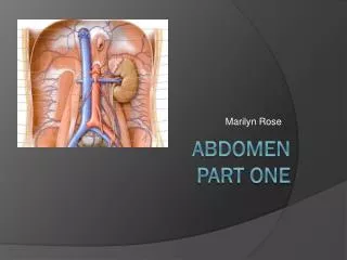
Abdomen Part 2 Marilyn Rose
Liver • Largest organ of abdomen • Rt hypochondriac/ and epigastric regions • Borders: • Superior/lateral and anterior= Rt diaph • Medial= sto/duodenum, transverse colon. • Inferior= hepatic flex • Posterior= Rt Kid • Glisson’s Capsule • Covered in peritoneum • Except: GB fossa, area of IVC and bare area.
Fissures of liver • Ligamentum teres- divides lt hepatic lobe into medial and lateral segments • Ligamentum venosum- separates the caudate lobe from Lt lobe • Transverse (portal) fissure- horizontal R/L PVS • Main lobar fissure (GB)- divides R/L lobes • Porta Hepatis- inferomedial border of the liver- the site of the MPV/HA and common bile duct.
Lobes of the liver • Lt lobe- • most anterior- lt of midline- separated from Rt by interlobar fissure/ Middle hepatic vein. • Caudate lobe • Smallest lobe, on the inferior/posterior surface, between the IVC and ligamentum venosum • Quadrate lobe- • Anterior-inferior surface of LT lobe between GB and ligamentum teres (remnant fetal umbilical vein, which runs along edge of falciform- The falciform supports/ attaches liver to diaphragm. 1: Right lobe of liver2: Left lobe of liver3: Quadrate lobe of liver4: Round ligament of liver5: Falciform ligament6: Caudate lobe of liver7: Inferior vena cava8: Common bile duct9: Hepatic artery10: Portal vein
Segments of Liver • Divided into 8 segments by the vascular supply (4=a/b) • Three hepatic veins: • MHV- divides liver into Rt/Lt lobes • RHV- divides Rt lobe into medial and lateral • LHV- Lt lobe into medial and lateral • Rt/ Lt Portal veins: • Divides each section transversely • Each segment is functionally independent with its own artery, portal vein and bile duct
Portal System • Nutrient rich blood from GI tract via the portal veins. • (75-80%) • Formed in the retroperitoneum by the superior mesenteric and splenic veins. • (posterior to the neck of the pancreas) • Porta hepatis- the portal vein branches into the Rt and Lt main portal veins- following the course of the HA’s • RPV branches into anterior and posterior branches • LPV courses left and then turns medially
End Stage Liver Failure Portal Hypertension-leads to Ascites and splenomegaly
Hepatic Artery • Arterial blood (20-25%) from common hepatic artery • Common hepatic artery arises from the celiac, entering the liver ANTERIOR to the portal vein. • The common hepatic artery arises from the celiac and branches into the Rt gastric and gastroduodenal arteries and continues ast eh proper heaptic towards the porta hepatis. • Prior to entering the liver it divides into the LT/RT hepatic arteries; which bring blood to each lobe.
Hepatic Veins IVC • Right Hepatic Vein • Largest- drains segments 5, 6 and 7 • Left Hepatic Vein • Smallest – drains segments 2,3 • Middle Hepatic Vein • Interlobar fissure- drains segments 4,5 and 8 • The three veins converge and enter the IVC just below the diaphragm
Biliary System • Gallbladder and bile ducts • Drain the liver and store bile until transported to the duodenum for digestion of fats.. • GB lives in the GB fossa- anterioinferior Rt lobe of the liver closest to the main lobar fissure. • Reservoir -fundus, body and neck- cystic duct Rt/Lt= CBD • Rt/Lt hepatic ducts unite at the porta hepatis to form the common hepatic duct (CHD), the CHD joins the cystic duct and forms the CBD. • CBD continues posterior to the pancreatic head and enters the duodenum along with the main pancreatic duct (Duct of Wirsung) at the Ampulla of Vater ( the muscle at the opening is called the sphincter of Oddi.
Biliary System Imaging ERCP MRCP Ultrasound
ERCP- Cholangiocarcinoma
Pancreas • Retroperitoneal, long, narrow- un-encapsulated • Posterior to the stomach/ between duodenum and splenic hilum • Head- located at the second portion of the duodenum about L2-L3, anterior to IVC and renal veins • Landmarks: • CBD Rt posterior and GDA on the anterior aspect • Uncinate process- between SMV and IVC • Neck- portal splenic confluence • main landmark- posterior to the neck • Body- largest, anterior to AO and SMA with the splenic vein running along the posterior surface • Tail- extends to the LT anterior pararenal space and LT kidney • Endocrine (insulin) and Exocrine (digestive enzymes) • Enzymes= amylase, lipase and peptidases and sodium bicarbonate
Spleen SAGITTAL • Largest lymph organ • Red pulp= blood • White pulp= lymphoid tissue and white blood cells • Intraperitoneal organ- covered in peritoneum • Posterior to stomach in the LUQ • Behind the 9-11th ribs • Borders- • medial- Lt kidney, splenic flexure, pancreatic tail • Posterior- diaphragm, pleura, Lt lung, ribs • Attached to > curvature of sto and Lt kid by the • Gastrosplenic and lienorenal ligaments TRANSVERSE
Spleen Splenic Rupture