Download
1 / 33
340 likes | 606 Vues
Principles of DNA and RNA Structure . PHAR 201/Bioinformatics I Philip E. Bourne Department of Pharmacology, UCSD Prerequisite Reading: Structural Bioinformatics Chapters 3 Thanks to Helen Berman for many slides. We start with DNA. History.

E N D
Principles of DNA and RNA Structure PHAR 201/Bioinformatics I Philip E. Bourne Department of Pharmacology, UCSD Prerequisite Reading: Structural Bioinformatics Chapters 3 Thanks to Helen Berman for many slides PHAR201 Lecture 2 2012
We start with DNA PHAR201 Lecture 2 2012
History • 1946 – DNA is the main constituent of genes (Avery) • 1950 – First X-ray pictures of DNA (Franklin) • 1953 – DNA structure revealed (Watson and Crick) • 1970 onwards - Multiple conformations and structures, initially from fibers • 1973 - X-ray structure confirms double helix (Rich) • 1974 - t-RNA structure (Kim) • 1980 – Structure of first complete turn of B (“normal”) DNA (Dickerson) PHAR201 Lecture 2 2012
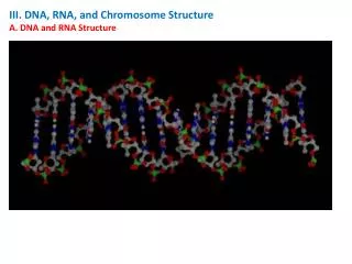
What Have we Learnt from These Structures? • Hydration, ionic strength and sequence all impact the type of structure • We see single stranded helices, double, triple and quadruple • Alone DNA and RNA does not crystallize easily, hence strands are short – eg 10-mer (unless complexed) • Contrast this to the ribosome (1FFK) PHAR201 Lecture 2 2012
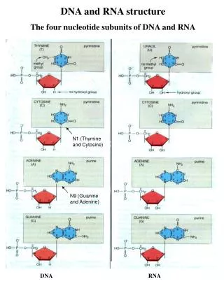
DNA and RNA Structure • NOTE: • Components • Sugar • Base • Phosphate • 5’ to 3’ direction • T->U in RNA • RNA - extra –OH at 2’ of pentose sugar • DNA - deoxyribose • Numbering • Single vs double strands • DNA more stable Voet, Donald and Judith G. Biochemistry. John Wiley & Sons, 1990, p. 792. PHAR201 Lecture 2 2012
NOTE: • Pyrimadines and Purines • T->U in RNA • Names • Numbering • Bonding character • Position of hydrogen • Tautomers Purines The 5 Basesof DNA and RNA Pyrimadines Neidle, Stephen. Nucleic Acid Structure and Recognition. Oxford University Press, 2002, p. 18. PHAR201 Lecture 2 2012
Tautomeric Structures • Keto vs enol (OH) • Different hydrogen bonding patterns Saenger, Wolfram. Principles of Nucleic Acid Structure. Springer-Verlag New York Inc., 1984, p. 113. PHAR201 Lecture 2 2012
Geometry of Watson Crick Base Pairs • A:T and G:C pairs are spatially similar • 3 H-bonds vs 2 (GC rich?) • Sugar groups are attached asymmetrically on the same side of the pair • Leads to a major and minor grove • Bases are flat but the hydrogen bonding leads to considerable flexibility • Base stacking is flexible Voet, Donald and Judith G. Biochemistry. John Wiley & Sons, 1990, p. 797. PHAR201 Lecture 2 2012
Definition of Major and Minor Groove Hydrogen bonding of WC base pair Mechanisms of recognition The canonical Watson-Crick base pair, shown as the G-C pair. Positions of the minor and major grooves are indicated. The glycosidic sugar-base bond is shown by the bold line; hydrogen bonding between the two bases is shown in dashed lines. PHAR201 Lecture 2 2012
Base Stacking is a Major Defining Feature of DNA Morphology • Dependant on: • Nature of the bases and base pairs • Stacking interactions • Explains sequence dependant features • Important for understanding molecular recognition PHAR201 Lecture 2 2012
Base Morphology The base-pair reference frame is constructed such that the x-axis points away from the (shaded) minor groove edge. Images illustrate positive values of the designated parameters. Reprinted with permission from Adenine Press from (Lu, et al., 1999). PHAR201 Lecture 2 2012
Backbone Conformation Voet, Donald and Judith G. Biochemistry. John Wiley & Sons, 1990, p. 807. PHAR201 Lecture 2 2012
A Beta-nucleoside • Ring is never flat – has 5 internal torsional angles • The pucker is determined by what is bound • A variety of puckers have been observed • Pucker has a strong influence on the overall conformation PHAR201 Lecture 2 2012
The Ribose Ring is Never Flat Voet, Donald and Judith G. Biochemistry. John Wiley & Sons, 1990, p. 808. PHAR201 Lecture 2 2012
The Glycosidic Bond Anti Syn • Connects ribose sugar to the base Neidle, Stephen. Nucleic Acid Structure and Recognition. Oxford University Press, 2002, p. 27. PHAR201 Lecture 2 2012
Change in sugar conformation affects the backbone C3’ C2’ C3’-Endo C2’ C3’ Voet, Donald and Judith G. Biochemistry. John Wiley & Sons, 1990, p. 808. C2’-Endo PHAR201 Lecture 2 2012
..and the position of the basesrelative to the helix axis A DNA B DNA PHAR201 Lecture 2 2012
Canonical B DNA Neidle, Stephen. Nucleic Acid Structure and Recognition. Oxford University Press, 2002, p. 34. PHAR201 Lecture 2 2012
Canonical B DNA • First determined experimentally by fiber diffraction (Arnott) • C2’-endo sugar puckers • High anti glycosidic angles • Right handed – 10 base pairs per turn • Bases perpendicular to the helix axis and stacked over the axis • Overall bending as much as 15 degrees (result of base morphologies – twist and roll) – {machine learning – sequence vs overall conformation?} • Over 230 structures 25 with base mis-pairing – only cause local perturbations • Strong influence of hydration along spine http://ndbserver.rutgers.edu/index.html PHAR201 Lecture 2 2012
Major Richer in base substituents Minor Hydrophobic H atoms of ribose groups forming its walls Major vs Minor Groove – distinctly different environments – important for recognition and binding PHAR201 Lecture 2 2012
Spine of Hydration Neidle, Stephen. Nucleic Acid Structure and Recognition. Oxford University Press, 2002, p. 97. PHAR201 Lecture 2 2012
A DNA Neidle, Stephen. Nucleic Acid Structure and Recognition. Oxford University Press, 2002, p. 36. PHAR201 Lecture 2 2012
Canonical A DNA Voet, Donald and Judith G. Biochemistry. John Wiley & Sons, 1990, p. 800. PHAR201 Lecture 2 2012
Canonical A DNA • C3’-endo sugar puckers – brings consecutive phosphates closer together 5.9A rather than 7.0 • Glycosidic angle from high anti to anti • Base pairs twisted and nearly 5A from helix axis • Helix rise 2.56A rather than 3.4A • Helix wider and 11 base pairs per repeat • Major groove now deep and narrow • Minor grove wide and very shallow PHAR201 Lecture 2 2012
Z-DNA • Helix has left-handed sense • Can be formed in vivo, given proper sequence and superhelical tension, but function remains obscure. • Narrower, more elongated helix than A or B. • Major "groove" not really groove • Narrow minor groove • Conformation favored by high salt concentrations, some base substitutions, but requires alternating purine-pyrimidine sequence. • N2-amino of G H-bonds to 5' PO: explains slow exchange of proton, need for G purine. • Base pairs nearly perpendicular to helix axis • GpC repeat, not single base-pair • P-P distances: vary for GpC and CpG • GpC stack: good base overlap • CpG: less overlap. • Zigzag backbone due to C sugar conformation compensating for G glycosidic bond conformation • Conformations: • G; syn, C2'-endo • C; anti, C3'-endo PHAR201 Lecture 2 2012
Z-DNA PHAR201 Lecture 2 2012
Z-DNA • Convex major groove • Deep minor groove • Alternate C then G • Spine of hydration PHAR201 Lecture 2 2012
Drug complexes to DNA • Bound to the base pair – double helix can accommodate this • Bound in the minor grove – show base specificity • Cis-platinum drugs PHAR201 Lecture 2 2012
Quadruplex DNA 1NP9 Jmol PHAR201 Lecture 2 2012
tRNA Invariant L-shape 1EVV jmol Saenger, Wolfram. Principles of Nucleic Acid Structure. Springer-Verlag New York Inc., 1984, p. 333. PHAR201 Lecture 2 2012
tRNA H bonds between distant regions Neidle, Stephen. Nucleic Acid Structure and Recognition. Oxford University Press, 2002, p. 148. PHAR201 Lecture 2 2012
The Ribosome • Complex of protein and RNA • Small 30S subunit – controls interactions between mRNA and tRNA • Large 50S subunit – peptide transfer and formation of the peptide bond PHAR201 Lecture 2 2012
Putting it all Together –Major Categories of DNA Binding Proteins Protein residues that make no contacts with the DNA are colored blue, those contacting the sugar-phosphate backbone are colored red, and those making base contacts are colored yellow. (a) Proteins with a single binding head: T4 endonuclease V (1vas), PU.1 ETS domain (1pue). (b) Proteins with a double binding head: lambda repressor (1lmb), papillomavirus-1 E2 DNA-binding domain (2bop). (c) Proteins with an enveloping mode of binding: NF-kB (1nfk),EcoRI restriction endonuclease (1eri). Jones et al. 1999 JMB 287(5) 877 PHAR201 Lecture 2 2012