Download
1 / 30
500 likes | 2.78k Vues
By: Nathan, Melissa, Shanik. Shoulder Joint (Glenohumeral Joint) . Surface Anatomy . Anterior axillary fold. Manubrium. Posterior axillary fold. Anterior axillary line. Surface Anatomy. Clavicle. Clavicular Deltoid origin. Clavipectoral triangle. Clavicular Pectoralis

E N D
By: Nathan, Melissa, Shanik Shoulder Joint (Glenohumeral Joint)
Surface Anatomy Anterior axillary fold Manubrium Posterior axillary fold Anterior axillary line
Surface Anatomy Clavicle Clavicular Deltoid origin Clavipectoral triangle Clavicular Pectoralis Major origin Sternocostal head of Pectoralis Major
Surface Anatomy Descending Trapezius Middle Trapezius Anterior Deltoid Acromial Deltoid origin Middle Deltoid Posterior Deltoid Ascending Trapezius Scapular Spine Deltoid origin Triangle of Auscultation
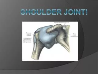
Classes of Joints There are three classes of joints in the body which are called: • Fibrous • Cartilaginous • Synovial The shoulder is a Synovial Joint
Types of Synovial Joints There are six types of synovial joints that occur in the body: • Plane or griddle Joints • Saddle Joints • Hinge Joints • Pivot Joints • Ball-and-socket Joints • Ellipsoid joints The shoulder is a ball-and-socket joint which allow for ROM in most directions
Types of Synovial Joints Plane Joint Saddle Joint Ball-and-Socket Joint
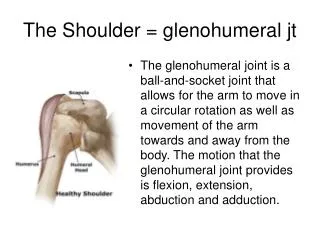
Movements of the Shoulder • The shoulder joint can do most ROM: • Flexion • Extension • Abduction • Adduction • Rotation • Circumduction
Synovial Membrane • Synovial joints are lined with a membranes called synovial membrane that secretes synovial fluid into the joint for : • Lubrication • Nourishes • Smooth movements • Filling all empty spaces
Articular Cartilage • Bone ends in the synovial joint are covered by hyaline cartilage called Articular Cartilage for smooth gliding movements • The humeral head articulates with the glenoid cavity of the scapula
Articular Capsule The articular cartilage is surrounded by a joint capsule made of: • Synovial Membrane • Fibrous Layer Helps hold the bones together and allows for movement to happen
Layers of Capsule • Fibrous layer aids in covering periosteum of the bone and helps with strength and stability of the joint • Synovial membrane covers the internal portion of the joint and secretes synovial fluid Capsule Membrane Fluid Articular Cartilage
Cartilage of Shoulder Because glenoid cavity is shallow, the head of humerus needs help to articulate with the cavity (Articular Cartilage on surface) GlenoidLabruim – ring of fibrocartilaginous material that attaches to the margin of the glenoid cavity FL GC SM GL FL- Fibrous Layer, GC- Glenoid Cavity, SM- Synovial Membrane, GL- GlenoidLabruim
Bursae • In some synovial joints, bursae are found • Extension of a synovial membrane that form into a sac • Filled with synovial fluid • Help cushion or protect tendons from rubbing against bones
Bursae of the Shoulder • Subacromial bursa or Subdeltoid bursa • Located between acromion, deltoid, and coracoacromial ligament • Helps with movement of supraspinatus tendon
Subscapular Bursa • Subscapular bursa or subcoracoid bursa • Located between the tendon of the subscapularis muscle and the neck and corocoidproccess of scapula • Protects and reduces friction between the tendon where it passes inferior to the coracoid process and over the neck of the scapula
Ligaments of the Shoulder Ligaments are fibrous tissue that connects bones to other bones. They are sometimes called articular ligaments • Coracohumeral • Transverse humeral • Coniod • Acromioclavicular • Glenohumeral • Coracoclavicular • Superior transverse scapular
Location of Ligaments • Acromiolclavicular ligament: Extends from the acromion to the clavicle • Coracoclavicular ligament: Anchors the clavicle to the coracoid process of scapula - Conoid: Attaches to the root of the coracoid process, base attaches to the inferior surface of the conoid tubercle of the clavicle • Glenohumeral ligaments: Part of the fibrous layer of the capsule. Consists of superior, middle, and inferior ligaments. All originate from the humerus to margin of glenoid cavity • Coracohumeral ligament: Root of the coracoid process to humeral neck • Transverse Humeral ligament: Broad fibrous band from greater to lesser tubercle. Holds the tendon from the long head of the biceps brachii muscle • Superior transverse scapular ligament: Attached by end of the coracoid process and inserts into the medial end of the scapular notch 1 2 4 6 5 3
Posterior Musculature Posterior Musculature Upper trapezius Levator scapulae Levator scapulae Middle Trapezius Middle Trapezius Rhomboid Minor Rhomboid Minor Infraspinatus Infraspinatus Rhomboid Major Rhomboid Major Posterior Deltoid Posterior Deltoid Teres Minor Teres Minor Teres Major Teres Major Serratus anterior Serratus anterior Lower trapezius
Posterior Musculature Trapezius Innervation: Spinal accessory nerve Vascularization: Transverse cervical artery Upper Action: Scapular elevation and upward rotation Middle A: Scapular retraction Lower A: Scapular depression and upward rotation Levator Scapulae I: 3rd and 4th Cervical nerves V: Dorsal Scapular Artery A: Scapular elevation and downward rotation Serratus anterior I: Long Thoracic Nerve V: Lateral Thoracic Artery A: Scapular protraction and upward rotation Rhomboideus Major and Minor I: Dorsal scapular nerve V: Dorsal scapular artery A: Scapular retraction and downward rotation
Posterior Musculature T1 Supraspinatus Supraspinatus Infraspinatus Deltoid Teres Minor Teres Major Latissimus Dorsi
Musculature Deltoids I: Axillary Nerve V: Posterior circumflex artery Anterior A: Shoulder flexion, medial rotation, horizontal adduction Middle A: Shoulder abduction Posterior A: Shoulder extension, hyperextension, lateral rotation, horizontal abduction Latissimusdorsi I: Thoracodorsal nerve V: Deep scapular artery A: Shoulder extension, adduction, medial rotation, hyperextension Teres Major I: Subscapular Nerve V: Circumflex scapular artery A: Shoulder extension, adduction, medial rotation
Anterior view of Musculature Manubrium Clavicle Subscapularis Pectoralis Major Coracobrachialis Anterior Deltoid
Musculature Pectoralis Major I: Lateral and Medial pectoral nerve V: Lateral Thoracic artery A: Shoulder adduction, medial rotation, horizontal adduction Pectoralis Minor I: Medial pectoral nerve V: Axillary artery A: Scapular depression, protraction, and downward rotation Coracobrachialis I: Musculocutaneous nerves C6, C7 V: Brachial artery A: Weakly adducts the shoulder joint Origin: Coracoid process Insertion: Medial aspect of humerus
Rotator Cuff The Rotator Cuff is made up of four muscles, Supraspinatus, Infraspinatus, Teres Minor and Subscapularis. The “SITS” muscles The tendons of these four muscles merge with the joint capsule of the shoulder as they pass it to insert on the tubercles of the humerus. This insertion forms a partial sleeve around the proximal end of the humerus. The Rotator Cuff reinforces the joint capsule and holds the head of the humerus in the glenoid cavity.
Muscles of the Rotator Cuff Subscapularis I: Subscapular nerve V: Subscapular artery A: Medial rotation Teres Minor I: Axillary nerve V: Circumflex scapular artery A: Lateral rotation, horizontal abduction Supraspinatus I: Subscapular nerve V: Subscapular artery A: Shoulder abduction Infraspinatus I: Subscapular nerve V: Subscapular artery A: Lateral rotation, horizontal abduction
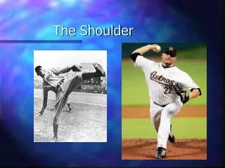
Rotator Cuff Tear Rotator Cuff Tear •Common in sports and recreation. •Joint is not protected ventrally •Supraspinatus is easily torn with: pitching (baseball) falls (skiing) hard blows from the side (hockey)