Download
1 / 66
730 likes | 1.4k Vues
GLENOHUMERAL JOINT (SHOULDER JOINT). Daniel Harris Amanda House Rebecca Miller Ginny Rinaldi. Ligaments. What is a ligament?.

E N D
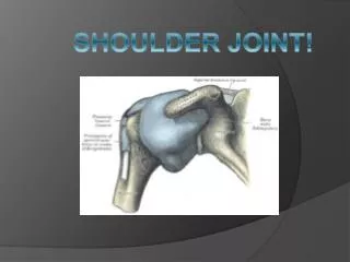
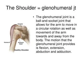
GLENOHUMERAL JOINT(SHOULDER JOINT) Daniel Harris Amanda House Rebecca Miller Ginny Rinaldi
What is a ligament? • A ligament most commonly refers to a band of tough, fibrous dense regular connective tissue that connects bones to other bones (not bones to muscles) to form a joint. Some ligaments limit the mobility of articulations, or prevent certain movements altogether. • Ligaments gradually lengthen when under tension, and return to their original shape when the tension is removed • The consequence of a broken/overstretched ligament can be instability of the joint. Over time instability of a joint can lead to wear of the cartilage which can eventually lead to osteoarthritis.
Acromioclavicular Ligament • The acromioclavicular ligament is at the top of the shoulder; it is part of the acromioclavicular joint, which lies between the acromion process and clavicle. The acromioclavicular ligament is divided into superior and inferior parts. • This ligament provides horizontal stability to the Acromioclavicular joint.
CORACOCLAVICULAR LIGAMENT • The coracoclavicular ligament is the combination of the conoid ligament, and the trapezoid ligament. Together these ligaments provide stabilization for the acromioclavicular joint. They are attached between the coracoid process of the scapula and the underside of the clavicle.
SUPERIOR TRANSVERSE SCAPULAR LIGAMENT • The superior transverse scapular ligament creates a small foramen from the scapular notch. This ligament is attached by one end to the base of the coracoid process, and by the other to the medial end of the scapular notch. • The ligament will sometimes ossify.
CORACOHUMERAL LIGAMENT • The coracohumeral ligament- a broad ligament which strengthens the upper part of the capsule of the shoulder joint. • It arises from the lateral border of the coracoid process, and passes diagonally downward and laterally to the front of the greater tubercle of the humerus. • Flexion, Extension
TRANSVERSE HUMERAL LIGAMENT • The transverse ligament of the humerus consists of a narrow sheet of connective tissue fibers that runs between the lesser and the greater tubercles of the humerus.Together with the intertubercular groove of the humerus, the ligament creates a canal through which the long head of the biceps brachi muscle passes.
GLENOHUMERAL LIGAMENTS • Three ligaments (seen in blue) on the anterior side of the glenohumeraljoint. Together these ligaments reinforce the anterior glenohumeral joint capsule. • The superior, middle, and inferior glenohumeral ligaments play different roles in the stability of the head of the humerus depending on arm position and degree of rotation. • Abduction, Adduction, External/Internal rotation
WHAT DO BURSAE DO? • Bursae are flattened sacs made of synovial membrane that are filled with synovial fluid. These sacs function as cushions between your bones and the muscles (deep bursae) or bones and tendons (superficial bursae) • Bursae reduce friction and allow your soft tissue to slide over bone easily during muscle contraction. • The synovial fluid found in bursae comes form synovial cells and are rich in protein and collagen. This acts as the lubricant between areas in your body where friction is greatest.
SUBDELTOID & SUBACROMIAL BURSAE • Subdeltoid bursa- Located between the deltoid muscle and the shoulder joint cavity and is usually joined to the subacromial bursa. • Subacromial bursa- Situated below the acromion process and above the greater tubercle of the humerus lessening the friction when you move your arm or raise it overhead.
SUBSCAPULAR & SUBCORACOID BURSAE • Subscapular bursa- Located between the joint capsule and the tendon of the subscapularis muscle. The subscapular bursa usually is continuous with the synovial cavity of the joint cavity. • Subcoracoid bursa- sits between the coracoid process of the scapula and the shoulder joint capsule. The grey arrow shows subcoracoid bursa
BURSITIS • When pressure or friction is too great, excess fluid can build up in the bursa sac causing swelling and inflammation. When a bursa becomes inflamed, moving the shoulder becomes very painful and movement can be difficult. Any actions that put pressure on the inflamed bursa can increase irritation and cause further inflammation and pain. • If the space becomes too crowded around the subacromial bursa, the acromion can begin to pinch the bursa or tendon causing an impingement (more commonly known as tennis shoulder) when your arm is raised in a forward reaching or overhead position.
CARTILAGE • Cartilage serves several functions, including providing a framework upon which bone deposition can begin and also supplying smooth surfaces for the movement of articulating bones. Cartilage is found in many places in the body and is classified as either "hyaline," "elastic," or "fibrous" cartilage. • Cartilage is distinctive in that it has only one cell type, is avascular (lacks blood vessels), aneural (no neurons and nerves), and alymphatic (no lymphatic system).
GLENOID LABRIUM • A fibrocartilaginous rim attached around the margin of the glenoid cavity in the shoulder blade. The glenoid fossa (the socket) of the scapula. Without the glenoid labrium only only one third of the head of the humerous would be covered (the ball) Therefore the socket is deepened by the glenoidal labrum.
ARTICULAR CARTILAGE • The term "articular cartilage" refers to the hyaline cartilage on the articular surfaces of bones. • Hyaline cartilage (aka “Gristle") is a type of cartilage found on many joint surfaces. It is pearly bluish in color with firm consistency and considerable collagen.
ARTICULAR CAPSULE • An articular capsule (or joint capsule) is an envelope surrounding a synovial joint. Each capsule consists of two layers: A fibrous layer and a synovail membrane. • On the inside of the capsule, articular cartilage covers the end surfaces of the bones that articulate within that joint. • The outer layer is highly innervated by the same nerves which go through through the adjacent muscles associated with the joint.
FIBROUS LAYER • An outer layer (fibrous strarum) of the articular capsule composed of avascular white fibrous tissue.
SYNOVIAL MEMBRANE • An inner layer (synovial stratum) of the articular capsule which is a secreting layer, and is usually described separately as the synovial membrane.
Rotator Cuff MusclesS.I.T.S • Supraspinatus • Infraspinatus
Teres Minor • Subscapularis
Clinical ConcernsR.O.M Normal Range of motion for the shoulder: • Abduction 180⁰ • Adduction 45⁰ • Extension 45⁰ • Flexion 90⁰ • Internal rotation 55⁰ • External rotation 40-45⁰
Rotator Cuff Injuries: • Rotator Cuff Conditions • Rotator cuff tear: An injury tears a rotator cuff tendon that’s been weakened by age or wear and tear. Weakness in the arm (and usually pain) are the symptoms. • Rotator cuff tendinitis (tendonitis): Repetitive overhead use of the arms (such as painting or throwing) causes a painful strain injury. Rest, ice, and pain relievers are usually effective treatments. • Rotator cuff impingement: The tendons of the rotator cuff are squeezed between the humerus and a nearby bone called the acromion. Symptoms and treatment of impingement are similar to tendinitis. • Frozen shoulder (adhesive capsulitis): The humerus adheres to the shoulder blade, causing shoulder pain and stiffness. Symptoms usually resolve with time and exercise, or steroid injections. • Subacromial bursitis: Inflammation of the small sac of fluid (bursa) that cushions the rotator cuff tendons from a nearby bone (the acromion).
Treatments • Pain medicines: Nonsteroidal anti-inflammatory drugs (NSAIDs), acetaminophen, or other medicines can be used to relieve the pain of rotator cuff injuries. • Corticosteroid injections: Cortisone or another anti-inflammatory steroid medicine is injected into the shoulder. The reduction in inflammation helps relieve pain. • Physical therapy: Various exercises can improve flexibility and strength of the other muscles in the rotator cuff. This increased strength can help compensate for a rotator cuff problem. • Occupational therapy: Similar to physical therapy, occupational therapy for rotator cuff injuries focuses on daily tasks that require shoulder movements. • Arthroscopic surgery: A surgeon operates through small incisions, using an arthroscope (a tube with a camera and tools on its end). The torn rotator cuff tendon is reattached to the bone. • Traditional (open) surgery: Through a larger incision, a surgeon cuts through the muscles and other tissues to reach a torn rotator cuff tendon. The tendon can then be reattached to the bone.
VEINS OF THE SHOULDER JOINT • CEPHALIC: The cephalic vein runs up the lateral side of the arm from the hand to the shoulder. In the shoulder, it pierces the tissues and empties into the axillary vein. After the cephalic vein joins the axillary vein, it becomes the subclavian vein and empties into the superior vena cava. • BASILIC: The basilic vein passes along the back of the forearm on the ulnar side for a distance and then curves to the surface below the elbow. It continues to move up the medial side until it reaches the middle of the upper arm. There, it enters deep into the tissues and joins the brachial vein. As the basilic and brachial veins merge, they form the axillary vein. • AXILLARY: The axillary vein is formed where the basilic and brachial veins come together, in the deep tissue of the upper arm.