Download
1 / 1
40 likes | 208 Vues
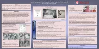
1933. 12 years. 1953. 17 years. 1970. 1991. Figure 1. Figure 2. Figure 3. Figure 4. Figure 5. Figure 7. Figure 8. MOHS MICROGRAPHIC SURGERY: A HISTORICAL PERSPECTIVE Patricia Ting, BSc, Anatoli Freiman, MD Division of Dermatology, McGill University Health Centre, Montreal, Canada.

E N D
1933 12 years 1953 17 years 1970 1991 Figure 1. Figure 2. Figure 3. Figure 4. Figure 5. Figure 7. Figure 8. MOHS MICROGRAPHIC SURGERY: A HISTORICAL PERSPECTIVE Patricia Ting, BSc, Anatoli Freiman, MD Division of Dermatology, McGill University Health Centre, Montreal, Canada ABSTRACT In the early 1930s, as a medical student at the University of Wisconsin, Fredric E. Mohs pioneered the idea of microscopically-controlled removal of cutaneous tumours. With its ability to systematically examine 100% of lesion margins, Mohs Micrographic Surgery (MMS) allows for maximal sparing of normal tissues and provides excellent cosmetic results. Whereas today, it is considered one of the greatest contributions to the field of dermatologic surgery, the widespread acceptance of the technique took over 50 years. We review the key historical events and people involved in the development of MMS. • INK DYES • Mohs refined his technique by color-coding the specimen edges with: • Red (merbromin) • Blue (laundry dye) • Black (India ink) • The addition of ink dyes provided a precise reference map of the tumour to be drawn and juxtaposed upon the corresponding surgical wound bed. Dr. Fredric E. Mohs (1910-2000) • THE BEGINNINGS • In the early 1930’s, as a young medical student at the University of Wisconsin, Frederic E. Mohs worked as an • assistant in a cancer research laboratory, investigating the effect of various irritants on implanted cancers in rats. • Mohs observed that injections of 20% zinc chloride effectively and accurately penetrated the tissues without • systemic toxicity. Furthermore, the chemical was also safe to handle, as it did not diffuse into the keratin layer of • the skin, unless a keratolytic, such as dichloroacetic acid, was previously applied. Most importantly, this zinc • preparation maintained the histologic architecture of tissues. • Mohs subsequently proposed the concept of chemical fixation followed by surgical excision of tumors with • microscopic examination of tumor margins. And thus, the concept of micrographic chemosurgery was born. • In 1936, after formal training as a general surgeon, Dr. Mohs began treating patients with cutaneous malignancies in • the dermatology clinic at the Wisconsin General Hospital. An elderly pharmacist at the drug store supplied him with • the ingredients required for the topical fixative paste containing stibinite (an antimony ore, which served as a • granular matrix) and sanguinaria canadenis (a binder for the saturated solution of zinc chloride). • FRESH TISSUE TECHNIQUE • In 1951, Dr. Ray Allington suggested that dichloroacetic acid could be used for hemostatis after the debulking • procedure. A monumental change in Mohs fixed tissue technique serendipitously occurred in 1953 during the filming of • an instructional documentary demonstrating Dr. Allington’s suggestion on a multistage removal of an eyelid basal cell • carcinoma. Due to the time constraints of the filming session, Mohs injected the residual tumour area with local • anesthetic and omitted the second round of the zinc chloride fixation. • It was soon realized that the resolution of tumor margins was not significantly impeded without fixation. This • became known as the fresh-tissue technique, which was also illustrated in Mohs’ chapter in the 1st edition of • Epstein’s Skin Surgery in 1956. • With this procedural modification, patients experienced significantly less pain and morbidity. Because of the • absence of tissue inflammation and sloughing, which were previously observed secondary to zinc chloride application, • more tissue could be conserved and reconstruction of the wound bed could now take place on the same day as the • excision. 1st Meeting of the American College of Chemosurgery in 1967 (Chicago). Front row & center: Dr. Fredric E. Mohs. Source: Brodland et al. 2000 • MILESTONES IN THE DEVELOPMENT OF MOHS MICROGRAPHIC SURGERY • 1933 Fredric Mohs’ original concept of chemosurgery for cutaneous neoplasms. • 1936 Dr. Mohs begins using his technique at the Wisconsin General Hospital. • Dr. Mohs reports 440 patients successfully treated with chemosurgery. • 1946 Mohs technique caught the attention of dermatologists at the American Academy of Dermatology meeting in Chicago. • The fresh tissue technique used for the first time during the filming of an instructional documentary on Mohs technique for the removal of eyelid tumours. • 1956 Mohs technique appears in the 1st edition of Epstein’s Skin Surgery. • Establishment of the first 1-year fellowship in Mohs surgery at NYU Medical Centre by Perry Robbins. • 1st American College of Chemosurgery meeting with 23 attendees. • 1969 Dr. Mohs presents a 100% cure rate for eyelid tumours using the fresh tissue technique at the 3rd American College of Chemosurgery meeting. • 1970 Dr. Theodore Tromovitch reports successful results with the fresh tissue technique at the 4th American College of Chemosurgery meeting. • Dr. Daniel Jones, a trainee of Dr. Mohs, coins the term “micrographic surgery." • 1st issue of the Journal of Dermatologic Surgery and Oncology published. • Textbook of Chemosurgery: Microscopically Controlled Surgery for Skin Cancer written by Dr. Mohs. • Minimum 1-year fellowships regulated by the American College of Chemosurgery. • American College of Chemosurgery renamed to American College of Mohs Micrographic Surgery and Cutaneous Oncology (ACMMSCO). • 1991 Establishment of the American Board of Mohs Micrographic Surgery and Cutaneous Oncology. • Adapted from Brodland et al. 2000 ORIGINAL PROCEDURE FOR MOHS CHEMOSURGERY – FIXED TISSUE TECHNIQUE (1936 – 1948) Step 1: Injection of local anesthetic (Figure 1) . Step 2: Removal and curettage of superficial tumour (Figure 2). Step 3: Application of dichloracetic acid (Figure 3) and in-situ zinc chloride fixative paste for 12 to 24 hours (Figure 4 & 5). Step 4: Surgical excision of tumour in saucer-like horizontal sections (Figure 6). Step 5: Microscopic examination of frozen sections. Figure 6. Diagrammatic representation of horizontal sections used in Mohs technique. • ACCEPTANCE OF MOHS MICROGRAPHIC TECHNIQUE • By the mid-1960s, Mohs technique gained wide acceptance, eventually leading to the establishment of American • College of Chemotherapy in 1967. • At the annual meeting in 1970, Dr. Theodore Tromovitch, who had been using the fresh tissue technique since the • 1960s, presented a case series of 75 patients treated for cutaneous carcinomas and reported equally high cure • rates with this new technique, which also proved that the real reason for the success of the fresh-tissue Mohs • surgery was not the chemical fixation of the tissue, but the microscopic control. • In 1986, the American College of Chemosurgery was renamed the American College of Mohs Micrographic Surgery • and Cutaneous Oncology (ACMMSCO) and minimum one-year fellowships in Mohs Micrographic Surgery were • officially regulated via this organization. • The Mohs surgery fellowship requires specialized training in a composite of skills including dermatopathology and • reconstructive surgery. Source: Mohs, FE. Chemosurgery – Microscopically Controlled Surgery for Skin Cancer (1978). • CONCLUSIONS • “Winning general acceptance takes much patience,” said Dr. Fredric Mohs. • The development and widespread acceptance of Mohs micrographic surgery has taken over 50 years. • Although the original concept of micrographic surgery began in the early 1930s, 12 years elapsed before the • development of the original fresh tissue technique (1936 to 1948), and its use for cutaneous carcinomas did not • begin until 17 years later (1953 to 1970). • With its ability to systematically examine 100% of tumour margins, Mohs Micrographic Surgery allows for maximal • sparing of normal tissues and provides excellent cosmetic results. • Today, it is considered one of the greatest contributions to the field of dermatologic surgery. INITIAL REJECTION OF MOHS FIXED TISSUE TECHNIQUE Between the 1930 – 1950s, Mohs’ pioneering surgical concept for the treatment of cutaneous malignancies was not well accepted by the medical community for several reasons: 1. Patients complained of extreme pain due to the caustic effects of zinc chloride fixative, which induced edema and tissue necrosis 2. Local inflammation made it difficult to interpret tumour histopathology 3. Microscopic examination of the margins and repeating process for incompletely excised tumor was very labor intensive 4. Increased infection rates due to delayed surgical reconstruction and closure of wound bed/defect 5. Belief that cutting through the tumour would promote local seeding of tumour cells and metastatic spread *Therefore, surgeons at the time preferred to use conventional wide surgical margins. • For residual neoplastic tissue, the process of fixation, surgical • excision and microscopic examination were repeated until the margins • cleared. • The defect was allowed to heal by secondary intention as the • fixative sloughed over the course of 7 to 10 days (Figure 7 & 8). • If required, skin grafts were performed after this time period. • * Mohs technique allowed for histological examination of entire base and • margins of the defect, ACKNOWLEDGEMENTS Special thanks to Dr. Lawrence Warshawski, Dr. Dan Issen & Dr. David Zloty (Vancouver, BC) for the clinical photos and to Dr. Channy Y. Muhn (Burlington, Ontario) for the inspiration. Source: Mohs, FE. Chemosurgery – Microscopically Controlled Surgery for Skin Cancer (1978).


![Applied Sampling [ Notes based on Graham Kalton’s Sage Publication and Prof. Jim Lepkowski’s Lecture Notes ]](https://cdn0.slideserve.com/487549/slide1-dt.jpg)