Download
1 / 55
550 likes | 697 Vues
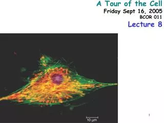
10 µm. A Tour of the Cell Friday Sept 16, 2005 BCOR 011 Lecture 8. Plasma Membrane – defines inside from outside. Common features of all cells. Outside of cell. Hydrophilic region. (a). Inside of cell. TEM of a plasma membrane. The plasma membrane, here in a red blood

E N D
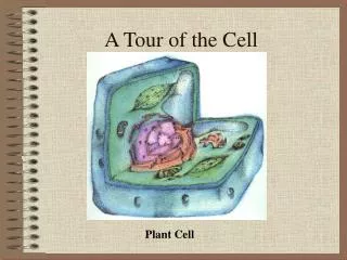
10 µm A Tour of the Cell Friday Sept 16, 2005 BCOR 011 Lecture 8
Plasma Membrane – defines inside from outside Common features of all cells
Outside of cell Hydrophilic region (a) Inside of cell TEM of a plasma membrane. The plasma membrane, here in a red blood cell, appears as a pair of dark bands separated by a light band. 0.1 µm Hydrophobic region Hydrophilic region Phospholipid Proteins (b) Structure of the plasma membrane Plasma membrane • Functions as a selective barrier • Specific portals for selective transport of materials in and out of cell Carbohydrate side chain Figure 6.8 A, B
Plasma Membrane – defines inside from outside Common features of all cells Cytosol - Semifluid “inside” of the cell • DNA “chromosomes” • - Genetic material – hereditary instructions Ribosomes – “factories” to synthesize proteins
Cytosol Free ribosomes ER • Carry out protein synthesis Membrane Bound ribosomes Proteins To be exported Large subunit Figure 6.11 TEM showing ER and ribosomes 0.5 µm Small subunit Ribosome – RNA & Protein Complex Diagram of a ribosome
Two Broad Classes of Cells Prokaryotes Eukaryotes Pro = before Eu = true karyon = nucleus HAVE A NUCLEUS membrane-bound organelles DO NOT HAVE A NUCLEUS NO internal membranes bacteria, cyanobacteria archaebacteria Plants, Animals, Fungi, protists
No internal membranes Bacterial Cell (Prokaryotic)
Pili: attachment structures on the surface of some prokaryotes Nucleoid: region where the cell’s DNA is located (not enclosed by a membrane) Ribosomes: organelles that synthesize proteins Plasma membrane: membrane enclosing the cytoplasm Cell wall: rigid structure outside the plasma membrane Capsule: jelly-like outer coating of many prokaryotes Bacterialchromosome 0.5 µm (a) A typical rod-shaped bacterium Flagella: locomotion organelles of some bacteria (b) A thin section through the bacterium Bacillus coagulans (TEM) Figure 6.6 A, B
On the same size scale: Bacterial cell (Prokaryotic Animal Cell (Eukaryotic)
Relative Sizes “Typical” ~ 1-2 M Bacterium “Typical” ~ 5 to 20 M diameter Animal Cell “Typical” ~ 5 to 50 M diameter Plant Cell M = micrometer or micron =10-6 meter
Internal membrane-bound organelles Animal Cell (Eukaryotic)
Why Internal Membranes? Compartmentalization (Division of Labor) I’m playing my sax I’m watching TV I’m cooking dinner I’m sleeping
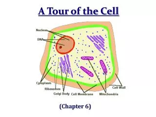
Animal Cell endoplasmic reticulum ENDOPLASMIC RETICULUM (ER) nucleus NUCLEUS Rough ER Smooth ER Plasma membrane cytosol Centrosome CYTOSKELETON Microfilaments Intermediate filaments ribosomes Ribosomes Microtubules Golgi apparatus Golgi apparatus Peroxisome lysosome In animal cells but not plant cells: Lysosomes Centrioles Flagella (in some plant sperm) Lysosome Figure 6.9 Mitochondrion mitochondrion
double membrane “Nuclear Envelope” nucleolus Nucleus: Information storage DNA housed, copied, read
The NUCLEUS Double membrane Nucleolus Nuclear pores DNA RNA protein lipid (membrane) Nuclear Lamina Euchromatin Heterochromatin
Nucleus Nucleus 1 µm Nucleolus Chromatin Nuclear envelope: Inner membrane Outer membrane Nuclear pores Pore complex Rough ER Surface of nuclear envelope. 1 µm Ribosome 0.25 µm Close-up of nuclear envelope Nuclear lamina (TEM). Pore complexes (TEM). nuclear envelope Figure 6.10 Nuclear lamina
Nucleolus Site of Ribosome Subunit Assembly Note: No membrane
Euchromatin region Site of mRNA synthesis Expression Of Informational RNAs
Endoplasmic reticulum (ER) • Smooth ER • Rough ER 1 m
Endoplasmic reticulum (ER) [Reticulum – network] Continuous network of flattened sacs tubules, vesicles, throughout eukaryotic cytoplasm Smooth ER • Synthesizes membrane lipids • Synthesizes steroids • Stores calcium • Detoxifies poison
Example: detoxification in smooth ER Benzo(a)pyrene charred meat, cigarette smoke Oxidations – more soluble Some metabolites are more toxic Chronic use of barbiturates, alcohol- SER proliferation, resistance
Rough ER – • ribosomes attached to cytoplasmic face • Large flattened sheets • Synthesizes secreted proteins, membrane proteinsexported • Protein modification; • initial steps of • carbohydrate addition • - glycoproteins
Rough ER Slips proteins Through ER membrane Glycosylation Adds oligosaccharides added as protein being made
1 Nuclear envelope is connected to rough ER, which is also continuous with smooth ER Nucleus Rough ER 2 Membranes and proteins produced by the ER flow in the form of transport vesicles to the Golgi Smooth ER cis Golgi 3 Golgi pinches off transport Vesicles and other vesicles that give rise to lysosomes and Vacuoles Plasma membrane trans Golgi 4 5 6 Lysosome available for fusion with another vesicle for digestion Transport vesicle carries proteins to plasma membrane for secretion Plasma membrane expands by fusion of vesicles; proteins are secreted from cell Figure 6.16
Golgi Apparatus: protein secretion Processing, packaging and sorting center Cis Golgi Close To RER Trans Golgi Far side Away From RER
Functions of the Golgi Apparatus cis Golgi - processing center “near” trans Golgi - sorting center “far” Present wrapping Service – modifies proteins Fed Ex Central Sorts for delivery To specific compartments
Functions of the Golgi Apparatus • Trimming of Oligosaccharide side chains on • glycosylated proteins • Addition of new Oligosaccharide residues to • existing side chains of glycosylated proteins • “Maturation” Cleavages of specific proteins • e.g., insulin • Phosphorylation of specific sugar residues on • oligosaccharide side chains of • glycosylated proteins • “molecular zip codes”
Molecular tags route proteins to proper destination P added in cis Golgi Proteins with M-6-P tag bind receptor in trans Golgi
Lysosomes: “Recycling Center” sacs of digestive enzymes
1 µm Nucleus Lysosome Hydrolytic enzymes digest food particles Food vacuole fuses with lysosome Lysosome contains active hydrolytic enzymes Digestive enzymes Lysosome Plasma membrane Digestion Food vacuole (a) Phagocytosis: lysosome digesting food Endocytosis And Phagocytosis Figure 6.14 A
EXTRACELLULAR FLUID 1 µm CYTOPLASM Pseudopodium Pseudopodium of amoeba “Food” or other particle Bacterium Food vacuole Food vacuole An amoeba engulfing a bacterium via phagocytosis (TEM). In pinocytosis, the cell “gulps” droplets of extracellular fluid into tiny vesicles. It is not the fluid itself that is needed by the cell, but the molecules dissolved in the droplet. Because any and all included solutes are taken into the cell, pinocytosisis nonspecific in the substances it transports. PINOCYTOSIS 0.5 µm Plasma membrane Pinocytosis vesicles forming (arrows) in a cell lining a small blood vessel (TEM). Vesicle In phagocytosis, a cell engulfs a particle by Wrapping pseudopodia around it and packaging it within a membrane- enclosed sac large enough to be classified as a vacuole. The particle is digested after the vacuole fuses with a lysosome containing hydrolytic enzymes. PHAGOCYTOSIS Figure 7.20
Receptor-mediated endocytosis enables the cell to acquire bulk quantities of specific substances, even though those substances may not be very concentrated in the extracellular fluid. Embedded in the membrane are proteins with specific receptor sites exposed to the extracellular fluid. The receptor proteins are usually already clustered in regions of the membrane called coated pits, which are lined on their cytoplasmic side by a fuzzy layer of coat proteins. Extracellular substances (ligands) bind to these receptors. When binding occurs, the coated pit forms a vesicle containing the ligand molecules. Notice that there are relatively more bound molecules (purple) inside the vesicle, other molecules (green) are also present. After this ingested material is liberated from the vesicle, the receptors are recycled to the plasma membrane by the same vesicle. RECEPTOR-MEDIATED ENDOCYTOSIS Coat protein Receptor Coated vesicle Coated pit Ligand A coated pit and a coated vesicle formed during receptor- mediated endocytosis (TEMs). Coat protein Plasma membrane 0.25 µm
Lysosome containing two damaged organelles 1 µ m Mitochondrion fragment Peroxisome fragment Lysosome fuses with vesicle containing damaged organelle Hydrolytic enzymes digest organelle components Lysosome Digestion Vesicle containing damaged mitochondrion (b) Autophagy: lysosome breaking down damaged organelle • Autophagy Figure 6.14 B
Vesicles move thru the endomembrane system exocytosis endocytosis
Mitochondria: Powerhouses of the cell
Mitochondria singular = mitochondrion • powerhouse of the animal cell • produces ~ 90% of ATP • Carries out oxidative reactions • Believed Derived from prokaryotic ancestor - DNA - ribosomes - double membrane – inner and outer *define two functional spaces
Mitochondrion Mitochondria are enclosed by two membranes • A smooth outer membrane • An inner membrane folded into cristae Intermembrane space Outer membrane Free ribosomes in the mitochondrial matrix Inner membrane Cristae Matrix Mitochondrial DNA Figure 6.17 100 µm
Cell – organelles = Cytosol Gel Important chemical reactions cytoskeleton - eukaryotes
Microtubule Microfilaments 0.25 µm Figure 6.20 • The cytoskeleton • Is a network of fibers extending throughout the cytoplasm • Structural Support • Movement of Materials and Organelles Figure 6.20
Microtubules Microfilaments Intermediate Filaments Table 6.1 Tubulin 25 mM dia Actin 7 mM dia various 8-15 mM dia Cell shape Organelle movt Chromosome separation Flagellar movt Cell shape Cell cleavage Cytoplasmic streaming Muscle contract Nuclear lamina Tension bearing elements Anchors There are three types of fibers that make up the cytoskeleton Motors: Dynein Kinesis Motors: Myosin
Vesicle ATP Receptor for motor protein Motor protein (ATP powered) Microtubule of cytoskeleton (a) Motor proteins that attach to receptors on organelles can “walk” the organelles along microtubules or, in some cases, microfilaments. Vesicles Microtubule 0.25 µm (b) Vesicles containing neurotransmitters migrate to the tips of nerve cell axons via the mechanism in (a). In this SEM of a squid giant axon, two vesicles can be seen moving along a microtubule. (A separate part of the experiment provided the evidence that they were in fact moving.) Figure 6.21 A, B • Movement of Vesicles along Microtubules
Motor MAPs transport vesicles Dynein inbound outbound kinesin MTOC
Centrosome Microtubule Centrioles 0.25 µm Longitudinal section of one centriole Cross section of the other centriole Microtubules Figure 6.22 • Contains a pair of centrioles • “microtubule-organizing center”
Animal cells • Lack cell walls • Are covered by an elaborate matrix, the ECM Polysaccharide molecule EXTRACELLULAR FLUID Collagen A proteoglycan complex Carbo- hydrates Core protein Fibronectin Proteoglycan molecule Plasma membrane Integrins CYTOPLASM Micro- filaments Integrin Figure 6.29 • The ECM Is made up of glycoproteins
Functions of the ECM include • Cell-Cell adhesion • Cell-Cell recognition • Regulation of cellular processes
Ribosomes (small brown dots) Rough endoplasmic reticulum Smooth endoplasmic reticulum NUCLEUS Golgi apparatus plant cell Central vacuole/Tonoplast Microfilaments Intermediate filaments CYTOSKELETON Microtubules Mitochondrion Peroxisome Plasma membrane Chloroplast Cell wall Wall of adjacent cell Plasmodesmata Figure 6.9
Central vacuole Cytosol Tonoplast Nucleus Central vacuole Cell wall Chloroplast 5 µm Plant Central vacuoles - Tonoplasts • Are found in plant cells • Hold reserves of important organic compounds and water • Regulates Turgor Figure 6.15
Chloroplast Ribosomes Stroma Chloroplast DNA Inner and outer membranes Granum 1 µm Thylakoid In plant cells, chloroplastscapture energy from the sun Photosynthesis Figure 6.18
Chloroplasts • Contain DNA • Contain bacterial-like ribosomes • Believed derived from prokaryotic ancestor • cyanobacterium = blue-green alga • -Double membrane organelle • defines three functional spaces
Inner Chlorplast Membrane 3 Central Players OuterChlorplast Membrane Stroma Thylakoid Space Intermembrane Space (transports things in and out of the chloroplast, but not central to photosynthesis itself Thylakoid Membrane