Download
1 / 31
310 likes | 322 Vues
Learn about the principles of spectroscopy and how it allows for precise measurements of energy differences between quantum levels. Explore atomic and molecular spectra and the information they provide. PowerPoints available for download.

E N D
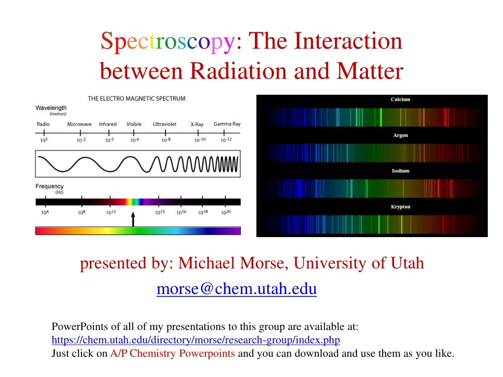
Spectroscopy: The Interaction between Radiation and Matter presented by: Michael Morse, University of Utahmorse@chem.utah.eduPowerPoints of all of my presentations to this group are available at: https://chem.utah.edu/directory/morse/research-group/index.phpJust click on A/P Chemistry Powerpointsand you can download and use them as you like.
The First Law of Spectroscopy: hν = Eupper state – Elower state and ν = c/λ (J-s·s-1 = J) (s-1 = m·s-1/m) Spectroscopy allows the differences between quantum energy levels to be measured to extraordinary precision, because the wavelength (λ) and the frequency (ν) of the light that causes the transition can be measured to exquisite precision. Only the energy differences can be measured, not the energy levels directly. Energy is measured in Joules. But more frequently spectroscopists use units proportional to energy, such as Hz (given by Even more commonly, cm-1 (wavenumbers) are used, denoted by upper state hν hν lower state Absorption Emission
The Electromagnetic Spectrum Covers a HUGE Range of Wavelengths Luckily, different types of excitations correspond (generally) to different wavelength (or frequency) regions. Excitation of rotational motions in molecules occurs in the microwave region: ν = 3×10⁹ to 3×10¹² s-1 or 3×10⁹ to 3×10¹² Hz λ = 10 cm to 0.01 cm ≡ 1/λ = 0.1 cm-1 to 100 cm-1 (cm-1 are called wavenumbers) ν and are proportional to the photon energy and are “quasi-energy units” Excitation of vibrational motions in molecules occurs in the infrared region: ν = 3×10¹² to 1.2×10¹⁴ s-1 (also known as Hz) λ = 100 μm to 2.5 μm = 100 cm-1 to 4000 cm-1 Excitation of electrons in atoms and molecules usually occurs in the near infrared, visible and ultraviolet regions: ν = 1.5×10¹⁴ to 3×10¹⁵ s-1 (also known as Hz) λ = 2000 nm to 100 nm = 5000 cm-1 to 100000 cm-1
Atomic Spectra: Excitation of Electrons Absorption Spectroscopy: Atoms are placed in the gas phase at relatively low temperature (maybe up to 5000K), and transitions from populated levels to higher lying levels are observed as absorption lines. This was first seen by Joseph von Fraunhofer (inventor of the spectroscope), who looked at the spectrum of the sun. Against a broad continuous emission spectrum, he observed sharp absorption lines that were due to atoms in the atmosphere surrounding the sun. Now we use atomic absorption spectroscopy as a quantitative technique for measuring the concentrations of specific atoms (Pb, Hg, Cd, etc.) in samples.
Atomic Spectra: Excitation of Electrons Emission Spectroscopy: In emission spectroscopy, atoms excited to high lying energy levels in an electric discharge or in a flame, and particular wavelengths of light are emitted that correspond to particular transitions. When examined with a spectroscope, these show up as sharp lines against a darker background. This technique can be used to examine the wavelengths emitted in flame tests for the various elements. This technique was developed extensively by Robert Bunsen (designer of the Bunsen burner) and Gustav Kirchhoff, who found samples that had previously unknown emission lines. In the process they discovered the elements rubidium and cesium. Emission spectrum of atomic cesium (Cs, NOT Cs+)
What can we learn from an atomic spectrum? Example: Aluminum atoms have a 1s2 2s2 2p6 3s2 3p1 configuration, where the spectrum is completely due to excitations of the 3p electron to higher lying ns or ndorbitals (excitations to other orbitals, like the np or nf orbitals, are forbidden):
What can we learn from an atomic spectrum? Zooming in on this higher energy range, the excitations get closer and closer as we approach the ionization limit:
What can we learn from an atomic spectrum? Extrapolation to the convergence limit of the series of lines lets up measure the ionization energy to extreme precision:
What about molecules? The most straightforward type of spectroscopy is pure rotational spectroscopy, in which the rotational motions are excited. For a diatomic molecule, the rotational energy levels are given by where J = 0, 1, 2, … is the rotational angular momentum quantum number, is the reduced mass, and R is the bond length. So analyzing a pure rotational spectrum allows the bond length to be measured! (In larger molecules, it allows the moments of inertia to be measured.) Defining (I is the moment of inertia), we get a simpler expression: . (B is called the rotation constant of the molecule) For nonlinear molecules, pure rotational spectra still exist but the energy levels depend on all three moments of inertia and the analysis is complicated. Still, this method gives highly precise geometrical information about small gas-phase molecules.
Pure rotational spectroscopy of linear molecules Usingwe get the energy level diagram: Because the photon that is absorbed has 1 unit of angular momentum, the rotational angular momentum, J, can only be increased by 1 unit, leading to a series of equally spaced absorption lines at energies of 2B, 4B, 6B, etc. Nonlinear molecules that have dipole moments also have pure rotational spectra – but they’re more complicated. 133Cs127I pure rotational spectrum (gas phase) B = 708.36 MHz R = 3.3151 Å To have a pure rotation spectrum, the molecule must have a dipole moment! The intensity of the spectrum is proportional to the dipole moment squared.
Pure rotational spectroscopy and astronomy How do we actually know anything about the composition of stars and outer space? One way: Radio Astronomy Rotating molecules in deep space emit radiation when they drop from one rotational state to another. This is in the radio or microwave portion of the spectrum, and can be detected using radio telescopes. Most known interstellar molecules have been detected by this method. Examples: H-C≡C-C≡C-C≡N H-C≡C-C≡C-C≡C-C≡N H-C≡C-C≡C-C≡C-C≡C-C≡N H-C≡C-C≡C-C≡C-C≡C-C≡C-C≡N H-C≡C-C≡C-C≡C-C≡C-C≡C-C≡C-C≡N FeO, NaI, SiC, SiN, SiO, SiS, TiO, TiO2 Many more
Where do you use rotational excitations in your everyday life?
Where do you use rotational excitations in your everyday life? Do you use a microwave oven? If so, you use rotational excitations to heat liquid water.
Where do you use rotational excitations in your everyday life? Do you use a microwave oven? If so, you use rotational excitations to heat liquid water. Microwaves of frequency ν=2450 MHz ( = 0.0817 cm-1 or λ=12.2 cm) are used to excite the hindered rotations of H2O in liquid water. Excitation of these motions quickly transfers to all other types of motion (hindered translations, etc.), heating liquid water. In ice, the H2O molecules are locked in place more strongly than in liquid H2O, so excitation of hindered rotations requires higher frequencies. The low frequency waves in a microwave oven don’t heat ice (at least not until a layer of liquid water forms on the surface, which then heats up quite effectively).
Microwave ovens are built to support 12.2 cm electromagnetic radiation Microwave ovens are built to support standing waves, where an integer number of half-wavelengths fit into the cavity. They reflect back and forth forming nodes where the electric field is always zero and crests where it oscillates positive and negative (just like a vibrating string). Guess what: Where there are nodes, your food doesn’t get hot! That’s why there’s a rotating platform so your food gets uniformly heated (more or less). Nodes, where the oscillating electric field is zero. At these spots, no microwaves can be absorbed.
How does light excite rotational motions? It’s all about the interaction between the dipole moment of the molecule and the electric field of the light. The interaction between a dipole moment () and the electric field ( causes a change in energy of the system given by If the dipole and the electric are properly aligned, the energy is lowered, as for the case of HF: Molecule favorably aligned Molecule unfavorably aligned +V +V F H F H -V -V
How does light excite rotational motions? If the molecule is oriented at an angle, the interaction energy can be lowered by rotating the molecule. In other words, the molecule feels a torque (a force that causes a rotation). By the time the molecule becomes aligned, the oscillating electric field has changed its phase, giving the molecule a force that drives it to align the opposite way. If the field oscillates at precisely the right frequency, the molecule can be rotationally excited. Molecule feels a torque +V F H -V
Molecules also have vibrations Diatomic molecules have vibrational energy levels labeled by the quantum number v, where v = 0, 1, 2, 3, … The v=0 level is the ground vibrational state. Typically, it is the only level that is significantly populated at room temperature. If the molecule’s dipole moment changes as the bond is lengthened or shortened (in other words, if ), then an electric field that oscillates at the right frequency can excite the vibrational motion of the molecule. The strongest absorption peak in the spectrum is the transition from v=0 to v=1 (the fundamental). Sometimes the v=0 to v=2 excitation (the first overtone) can also be observed. These transitions occur in the infrared part of the spectrum.
How does light excite vibrational motions? Again, it’s all about the interaction between the dipole moment of the molecule and the electric field of the light. Now, however, μ is a function of the bond length, R. If the dipole and the electric are favorably aligned, and the dipole moment increases when R increases, the molecule can lower its energy by stretching, which increasesto increase. When the electric field reverses direction, the force on the molecule reverses, causing R to decrease. If the oscillatory frequency of the field matches the vibrational frequency of the molecule, its vibrational motion can be excited. Molecule favorably aligned Molecule stretches in response +V +V F F H H -V -V
Some molecules can’t absorb IR light at all Some molecules have electric dipole moments that are zero at all bond lengths. These are molecules like H2, N2, and O2 that can never develop a dipole moment at any internuclear separation. For them is always 0. This is the reason why N2 and O2 aren’t greenhouse gases. They don’t absorb in the infrared, so IR light radiating from the warm earth passes right through them. H2O vapor, CO2, and CH4 do absorb IR light and re-radiate it, trapping heat that would otherwise escape into space.
Vibrations of polyatomic molecules If you imagine a molecule to be a set of masses (the atoms) connected by springs, you can solve Newton’s equations to find certain ways the molecule can vibrate in which all the atoms oscillate about their equilibrium positions with the same frequency. These are called normal modes of vibration. All solid objects have these normal modes – they’re why a glass vibrates with a particular frequency when we tap it will a fork or why a tuning fork can be designed to vibrate at a particular frequency. Even bridges have normal modes of vibration.
Polyatomic molecules have many normal modes A useful way to think about a polyatomic molecule with N atoms is this: To specify the location of all N atoms, you need 3N coordinates: (x1, y1, z1) for atom 1, (x2, y2, z2) for atom 2, etc. These 3N coordinates can be recombined to give (X, Y, Z) for the location of the center of mass and then 3N-3 other coordinates are needed to specify the locations of the atoms. Motion along the (X, Y, Z) coordinates gives translation of the molecule in space. If the molecule is linear, 2 angles are needed (θ, φ) to specify the position of the molecular axis. Motion in these coordinates is molecular rotation. That leaves 3N-5 coordinates to describe the position of the atoms relative to the rotating coordinate system. Movement along these coordinates gives the vibrations of the molecule. If the molecule is nonlinear, 3 angles are needed to specify the orientation of the molecule in space (3 rotational degrees of freedom). This leaves 3N-6 vibrations. Overall, linear molecules have 3N-5 different vibrational motions (normal modes); Nonlinear molecules have 3N-6 different vibrational motions (normal modes). CO2 has 4 normal modes H-C≡C-H has 7 normal modes C6H6 has 30 normal modes
Some normal modes can be excited by infrared radiation If the dipole moment of the molecule changes as it vibrates in a particular normal mode, that mode can be excited by IR light of the correct frequency. For example, CO2: Bending mode 667 cm-1 Symmetric stretch 1388 cm-1 Antisymmetric stretch 2349 cm-1 Dipole moment changes as the molecule bends. This mode is IR active Dipole moment remains zero as the molecule symmetrically stretches. This mode is NOT IR active Dipole moment changes as the molecule stretches antisymmetrically. This mode is IR active We expect IR absorptions at 667 cm-1 and 2349 cm-1, but NOT at 1388 cm-1. Is this what is observed?
The infrared spectrum of air CO2 antisymmetric stretch at 2349 cm-1. OBSERVED CO2 symmetric stretch at 1388 cm-1. NOT OBSERVED CO2 bending vibration at 667 cm-1. OBSERVED Spectrum is dominated by CO2 and H2O; N2, O2, and the symmetric stretch of CO2 are not observed.
Infrared spectra help to identify molecules C-H stretch C≡N stretch benzonitrile C-H stretch C=O stretch O-H stretch 4’-hydroxyacetophenone
Electrons in molecules can also be excited Here the excited state is less strongly bound than the ground state, so the well isn’t as deep. Also, the equilibrium bond length is a little longer. This could result from exciting a bonding electron to an antibonding orbital. Different wavelengths can be absorbed to take the molecule from a particular vibrational level, v”, in the lower state to a particular level, v’, in the excited state. If all the molecules are in v”=0, then a simple series of absorption bands are observed. These are labeled the v’-v” bands. A + B* Vibrational levels are labeled by v’ in excited state Excited electronic state: one electron has been excited to a higher energy molecular orbital AB* hν A + B Vibrational levels are labeled by v” in ground state Ground electronic state v” = 2 v” = 1 AB v” = 0 Upper state vLower state v
Spectrum of gaseous I2 at low temperature (simulated) Usually, spectra aren’t this clean and easily interpreted. You can tell, though, that useful information can be obtained – like the bond energy and vibrational frequency.
In a polyatomic molecule, more than one normal mode can be active • + vibrational energy for mode 6 vibrational energy for mode 6 vibrational energy for mode 5
Electronic emission spectroscopy of diatomic molecules When a molecule is excited in an electric discharge (like N2 in Prof. Statler’s discharge experiment), the excited state molecules can fluoresce to many different vibrational levels of the lower state. Then we see a series of nearly equally spaced emission bands. If several vibrational levels are populated in the excited state, we can get several series of emission bands (as in N2 below). A + B* Vibrational levels are labeled by v’ in excited state Excited electronic state v’=0 level AB* A + B Vibrational levels are labeled by v” in ground state A series of transitions from v’=0 to various v” levels shows up as a series of bands that are nearly equally spaced. Lowerelectronic state AB
Thanks for listening! Anytime you’d like to ask me a question: morse@chem.utah.edu This and all previous presentations can be found at my website: https://chem.utah.edu/directory/morse/research-group/ap_chemistry_powerpoints.php


