Download
1 / 29
290 likes | 319 Vues
Explore the impact of aging on the brain, including volume reduction, nerve cell changes, and microscopy findings such as lipofuscin increase and neurodegenerative markers. Learn about diseases like Alzheimer's, Pick's, Progressive Supranuclear Palsy, and Parkinson's, their onset, progression, and characteristic features.

E N D
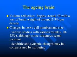
The ageing brain • Volume reduction: begins around 50 with a loss of brain weight of around 2-3% per decade • Changes in nerve cell numbers and size - various studies with various results ( 10-25%), although some structures seem resistent - dendritic and synaptic changes may be compensated by sprouting
Microscopy of the aging brain • Increase in lipofuscin • Senile plaques • Neurofibrillary tangles • Granulovacuolar degeneration • Hirano bodies • Leukoaraiosis • Amyloid (congophilic) angiopathy
Neurodegenerations • Damage to lysosomes, nuclear DNA, mitochondria leads to release of caspases, initiating apoptosis. • In many if not most neurodegenerations this damage is started by abnormal protein-protein interactions leading to protein storage. • Examples: • Ubiquinopathies • Tauopathies • Synucleinopathies • Prion diseases
Ubiquinopathies: Alzheimer’s disease Pre-senile and senile forms Onset before or after 65 years of age Progression slow (often > 1 decade) Pathological changes are similar to aging, but more severe
Senile plaque Heterogeneous structure Amyloid core surrounded by a clear zone Crown of filamentous or granular material made up of neuronal projections filled with argyrophilic filaments and debris Macrophages and astrocytes
Tauopathies • Pick’s disease • Progressive Supranuclear Palsy • Cortico basal degeneration • etc
Pick’s disease • Dementia with relative rapid progression with frontal symptoms (aggressive behavior often, or problems with initiation of tasks), onset from 50 years of age, frequently hereditary. • Gross findings are characterized by a lobar atrophy affecting the frontal lobe and the anterior temporal lobe (mostly the anterior 1/3 of the superior temporal gyrus) • Histology:Pick’s cells (swollen neurons ), Pick’s bodies ( argyrophilic intracytoplasmic inclusions), neuronal loss and gliosis
Progressive supranuclear palsy • Dementia combined with vertical gaze paralysis, and Parkinsonism • Abnormal tau accumulation in neurons and glial cells, neuronal loss and gliosis • Characteristic topography:In the midbrain and striatum (Substantia nigra, red nucleus, colliculi, globus pallidus, subthalamic nucleus, periaqueductal grey matter, dorsal and medial raphe, locus coeruleus)
Synucleinopathies • Parkinson’s disease • Dementia with Lewy bodies • etc
M. Parkinson • Onset 30-80 • Rest tremor mostly starting with hands (like counting money) • Stooped posture, walking with small steps • Depigmentation of Substantia nigra and Locus coeruleus • Lewy bodies and loss of pigmented neurons • Loss of dopaminergic innervation to the striatum