Plasmolysis Lab
Plasmolysis Lab. Plasmolysis . Plasmolysis is the loss of water from the cell by osmosis, and this is evident when the cell contents pull away from the rigid cell wall as the water moves out . Steps of Plasmolysis. TIME, ( min). Steps of Plasmolysis. Plasmolysis.

Plasmolysis Lab
E N D
Presentation Transcript
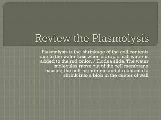
Plasmolysis Plasmolysis is the loss of water from the cell by osmosis, and this is evident when the cell contents pull away from the rigid cell wall as the water moves out.
Steps of Plasmolysis TIME, (min)
Plasmolysis Purpose: The purpose of this lab experiment is to demonstrate a biological principle observed in plant cells called plasmolysis. Plasmolysis is the loss of water from the cell by osmosis, and this is evident when the cell contents pull away from the rigid cell wall as the water moves out. Materials and Methods: For this experiment, the student requires a microscope, clean slides and cover slips, dropper, salt, spatula, and elodea. Prepare a wet mount using a single leaf. Carefully add some salt crystals to the edge of the cover slip. You may need to add a drop more water onto the crystals with the dropper. Now place the slide on the microscope stage and observe the leaf cells on low and then high power. Experiment: As water moves out of the cells by osmosis, you should observe the cytoplasmic contents clumping away from the cell wall as shown in the diagram below. Label the cell wall, central vacuole, cell membrane, and cytoplasm on the cell shown below at left.
In the diagram below, the cell is seen under normal conditions. The cell is at Dynamic Equilibrium! LC HC HC LC Field of View is 500um. So the cell is _____um?
In the diagram below, a 6 % salt solutions was added. This cell was viewed after five minutes. Plasmolyzed cell HC LC • Field of View is 500um. So the cell is _____um? • Did the entire cell get smaller or did • the contents shrink?
In the diagram below, the cells’ environment has been re-established using distilled water. LC HC Field of View is 500um. So the cell is _____um?
In this experiment what is considered hypertonic? What is considered hypotonic? In the diagram above, the right cell is plasmolyzed. The cell on the left is turgid. What does this mean and under what conditions would it be in this condition? Conclusion:
Plasmolysis of human blood cells after Addition of Salt Epidermis Cells of Allium cepa and Rhoeo discolor. Epidermis cells with stained vacuoles (red) are especially well suited for the depiction of plasmolysis.