Download
1 / 32
320 likes | 602 Vues
Other Systems in the Mammary Gland: Support, Nervous, Circulatory, and Lymphatic. Melissa Hlavacek. Support Systems. Variation across species. Litter-bearing Species. No large suspensory ligaments Fibrous connective tissue and skin are the primary support

E N D
Other Systems in the Mammary Gland: Support, Nervous, Circulatory, and Lymphatic Melissa Hlavacek
Support Systems • Variation across species
Litter-bearing Species • No large suspensory ligaments • Fibrous connective tissue and skin are the primary support • Examples: pigs, dogs, cats, rodents
Humans and Litter-Bearing Species • Gland is on top of muscle tissue • Strong, fibrous connective tissue separates it from the muscle • Suspensory ligaments are either attached to the muscle or the connective tissue • In litter bearing species, each half is also separated from the other • Midline • Skin also provides protection and a little support
The Cow • Many different tissues… * Median suspensory ligament * Skin * Lateral suspensory ligament - Superficial fascia - Fine connective tissue - Subpelvic tendon - Coarse connective tissue © The Babcock Institute
Connective Tissue & Skin • Fine connective tissue • Attaches skin to underlying tissue • Coarse connective tissue • Attaches front quarters to abdominal wall • Skin • Little support • Protection from pathogens • Front and rear are also separated by connective tissue • No internal crossover between any of the quarters
Median Suspensory Ligament • Primary support of the udder • Two adjacent heavy sheets of tissue • Mostly elastic, some fibrous tissue • Attaches to the abdominal wall • Divides the udder into halves (left and right) • Glands on each half are divided by sheets of tissue © Biology of Lactation, Schmidt
Lateral Suspensory Ligaments • Like a “hammock” around the udder • From the pelvis to the median suspensory ligament • Mostly fibrous tissue • Collagen • Attaches to the alveolar tissue • Provides internal framework • Does not connect at the base of the udder
How much support is enough? • High producing Holstein cow • Empty Udder = 50 lb. • Milk = 60 lb. • 50 + 60 = 110 lb. !!!
Importance • Udder support can significantly affect milking ability and useful life • 10 – 25% heritable • Pendulous udders are more likely to suffer injury • Especially teats • Increased mastitis • Affect on offspring
Nervous System • Few nerves go into the gland • Like other skin glands…no parasympathetic innervation
Sensory Nerves • Skin and teats • Positive stimulation of teats and surrounding area initiates milk let-down reflex via oxytocin • Critical! © Biology of Lactation, Schmidt
Sympathetic Nerves • Associated with arteries in the gland • Control blood flow to the gland • Carry oxytocin to the gland • Innervation of sphincters muscles in teats • Stress causes vasoconstriction decreasing milk secretion and let-down • There are no nerves to myoepithelial cells or alveolar cells • Contraction is regulated by oxytocin
Nervous System • In species such as pigs, nerve supply to the abdominal mammary glands is different than inguinal mammary glands.
Circulatory System • All milk precursors come from the blood • No crossover between each side of the mammary gland • Venous circle • Prevents pinching off of areas of venous outflow when animal is lying down
Circulation • Elongation and proliferation of growing vessels • Angiogenesis • Vasculogenesis • Complex interactions between • Endothelial cells • Extracellular matrix • Specific stromal cells • Requires dramatic reorganization of surrounding tissue
Growth factors affect rates of endothelial cell proliferation and degree of vessel formation • VEGF-vascular endothelial growth factor • Major regulator • Maintains viability • Stimulates mitogenesis and chemotaxis • Changes permeability • Many growth factors in different isoforms • Enzymes alter surrounding tissue
Sphincters • Capillary beds • alveolus ‘hairnet’ • Sphincters constricted • no blood flow • Sphincters open • blood flow to alveolus
Local control of blood flow • High pO2 = high blood flow • Need ATP • ATP needs oxygen • High pO2= high ATP • = sphincters constricted Terminal arteriole Metarteriole Large vein sphincters Post capillary venule
Other local controllers of blood flow * Oxygen * Carbon Dioxide • Adenosine • Lactic Acid • pH • Hormones • Nitric Oxide • Alveolar cells each control their own blood and nutrient supply
So how much blood is enough? 500 Liters blood = 1 Liter milk 1 truck of blood = 100 lbs. milk
Hot area of research: mammary tumors Depends on angiogenesis for survival Killer of females and males
Estrogen • Mediates synthesis/secretion of local tissue growth factors • Direct effect? • Changes in angiogenesis of ovary, placenta, endometrium • Mammary gland is a reproductive organ • Other hormones?
Tumors • What’s u-PA? • Activator of the zymogen plasminogen • Plasmin degrades extracellular proteins • Increases angiogenesis and mammary development • High concentrations of u-PA in breast cancer patients
Thermogenesis Hot spot Infra-red thermography
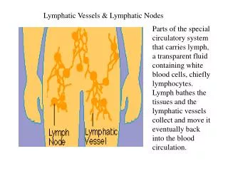
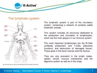
Lymphatic System • Molecules go out of the capillaries but not all can get back in • Disrupt the normal osmotic pressure • Functions • Pick up extracellular fluids and bring back to the circulatory system • Transport of leukocytes • Transport of immune cells
Lymphatic System • 2 Supramammary Lymph Nodes • Each above ½ the udder • Filter tissue fluids • Remove foreign material • Vaccination?
Udder Edema • Parturition • Accumulation of secretions • Pressure • Passive flow is hindered • Tight junctions do not function well • Fluid enters interstitial space • Alters osmolarity • Alters hydrostatic pressure • Less fluid is removed • Connective tissue spaces swell • Causes???