Chronic Renal Failure
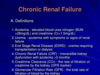
Chronic Renal Failure. Definition. Kidney damage for more than 3 months, as: structural or functional abnormality of the kidney with or without decreased GFR manifested by either pathologic abnormalities or markers of kidney damage, including:

Chronic Renal Failure
E N D
Presentation Transcript
Definition • Kidney damage for more than 3 months, as: • structural or functional abnormality of the kidney • with or without decreased GFR • manifested by either pathologic abnormalities or markers of kidney damage, including: • abnormalities in the composition of blood or urine or abnormalities in imaging tests • Glomerular filtration rate less than 60 ml/minute/1.73 m2 for 3 months, • with or without kidney damage
Stagesof CKD • Stage 1 • kidney damage with normal or increased GFR (90 or more rnL/minute/1.73m2) • Stage 2 • kidney damage with mild decrease in GFR (60-89 ml/minute/1.73m2 • Stage 3 • moderate decrease in GFR (30-59 mL/minute/1.73 m2) • Stage 4 • severe decrease in GFR (15-29 ml/minute/1.73 m2) • Stage 5 • kidney failure (less than 15 mL/minute/1.73 m2 or on dialysis
Etiology • Diabetes (40% of new cases of ESRD in the United States) • Hypertension (25% of new cases) • Glomerulonephritis (10%) • Others-urinary tract disease, polycystic kidney disease, lupus, analgesic nephropathy, unknown
RiskFactors • Susceptibility • associated with an increased risk, but not proved to cause CKD: • Advanced age, reduced kidney mass, low birth weight, racial/ethnic minority, family history, low income or education, systemic inflammation, and dyslipidemia. • Mostly not modifiable • Initiation • directly cause CKD: • diabetes, hypertension, autoimmune disease, polycystic, kidney diseases, and drug toxicity. • Modifiable by drug therapy • Progression • result in faster decline in kidney function: • hyperglycemia, elevated BP, proteinuria, and smoking
Albuminuria/Proteinuria • Marker of kidney damage, progression factor, and cardiovascular risk factor. • Can be classified follows • Normal: albumin excretion less than 30 mg/24 hours • Microalbuminuria: 30-300 mg/24 hours • Macroalbuminuria: (overt proteinuria) more than 300 mg/24 hours • Nephroticrange proteinuria: more than 3 g/24 hours • Assessment for proteinuria • most commonly assessed by measurement of urinary albumin, creatinine ratio. • Spot urine: Untimed sample is adequate for adults and children (screening test).
AssessmentofKidneyFunction • Serum creatinine • Avoid use as the sole assessment of kidney function. • Dependent on age, sex, weight, and muscle mass • Many laboratories use "standardized" Cr traceable to IDMS (isotope dilution mass spectrometry). • This will alter calculations (Cockcroft-Gault and MDRD). Make sure you know if the Cr is standardized. • Measurement of GFR. Inulin, iothalamate, and others are not routinely used. • Measurement of CrCl through urine collection • Reserve for vegetarians, patients with low muscle mass, patients with amputations, and patients needing dietary assessment, as well as when documenting need to start dialysis. • Urine collection will give a better estimate in patients with very low muscle mass. In most cases, equations will overestimate kidney function because Cr concentrations will be low in patients with very low muscle mass.
AssessmentofKidneyFunction • Calculated using Cockcroft-Gault (mL/minute CrCl) - overestimates GFR [(140- age) x body weight]/[SCr x 72] x (0.85 if female) • Estimated GFR with MDRD study data • Estimated GFR (mL/minute/1.73 m2) in patients with known CKD (less than 90 mL/ minute) • Abbreviated MDRD formula correlates well with the original MDRD formula, simpler to use GFR (mL/minute/1.73 m2) 186 X scr-1.154 X age-0·203 X (0.742 if female) X (1.21 if African American). • This equation and an adjusted equation for use with standardized creatinine are available at www.nkdep.nih.gov or www.kidney.org. • CKD-EPI (Chronic Kidney Disease Epidemiology Collaboration) equation. Alternative equation to estimate GFR. See www.kidney.org. • For children, Schwartz and Counahan-Barratt formulas
DiabeticNephropathy • Pathogenesis • Hypertension (systemic and iutraglomerular) • Glycosylation of glomerular proteins • Genetic links • Diagnosis • Long history of diabetes • Proteinuria • Retinopathy (suggests microvascular disease) • Monitoring • Type I-Begin annual monitoring for microalbuminuria • 5 years after diagnosis. • Type II-Begin annual monitoring for proteinuria immediately • (do not know how long they have had diabetes mellitus).
DiabeticNephropathy • Management/slowing progression • Aggressive BP management • Target BP in patients with diabetes and CKD is less than 130/80 mm Hg. • ACEI's and ARBs are preferred • often in combination with a diuretic • should be used with any degree of proteinuria, even if the patient is not hypertensive. • Calcium channel blockers • nondihydropyridineare 2nd line to ACE/ARBs. • Data are emerging for combined therapy. • Intensive blood glucose control. Glycosylated hernoglobiu less than 7%. Less aggressive with more advanced CKD • Protein restriction-There are insufficient data in diabetes. Patients should avoid high protein diets.
NondiabeticNephropathy • Manage hypertension. • If proteinuricand hypertensive, • use ACE and ARB. • Minimize protein in diet. • Controversial. • May slow progression based on MDRD study but may also impair nutrition
Other Guidelines to Slow Progression • Manage hyperlipidemia. • Follow National Cholesterol Education Program guidelines. • Goal is low-density lipoprotein (LDL) less than 100. • Statins are first line. • Stop smoking.
RENAL REPLACEMENTTHERAPY • Indications for Renal Replacement Therapy A- acidosis • not responsive to bicarbonate) E- electrolyte abnormality • hyperkalemia; hyperphosphatemia I- intoxication • boric acid; ethylene glycol; lithium; methanol; phenobarbital; salicylate; theophylline) 0- fluid overload • symptomatic [pulmonary edema]) U- uremia • pericarditis and weight loss
RENAL REPLACEMENTTHERAPY • Two Primary Modes of Dialysis • Hemodialysis-most common modality • Peritoneal dialysis
Hemodialysis Access (intermittent for ESRD) • Arteriovenousfistula-preferred access! • Natural, formed by anastomosis of artery and vein • Lowest incidence of infection and thrombosis, lowest cost, longest survival • Takes weeks/months to "mature“
HD Access • Arteriovenous graft • Synthetic (polytetrafluoroethylene) • Often used in patients with vascular disease
HD Access • Catheters • Commonly used if permanent access not available • Problems include high infection and thrombosis rates. Low bloodflows lead to inadequate dialysis.
HD • Dialysis membranes • Conventional • not used much anymore. Small pores. Made of cuprophane • High flux and high efficiency • large pores. • Can remove drugs that were impermeable to standard membranes (vancomycin). • Large amounts of fluid removal (ultrafiltrate)
HD • Adequacy • Kt/V-unitlessparameter. • K=clearance, t=time on dialysis, and V=volume of distribution of urea. • Kidney Disease Outcomes Quality Initiative set goal of 1.2 or more. • URR-urea reduction ratio. • URR=BUN post/BUN pre x 100%. • Goal is URR greater than 65%
HD • Common complications of HD a. Intradialytic • Hypotension-primarily related to fluid removal. Common in people who are elderly and people with diabetes mellitus. Tx: limit fluid gains between sessions; give normal or hypertonic saline, midodrine. Less well-studied agents include fludrocortisone, selective serotonin reuptake inhibitors • Cramps-vitamin E or quinine (controversial because of adverse effect profile) • Nausea/vomiting • Headache/chest pain/back pain b. Vascular access complications-most common with catheters • Infection-S. aureus. • Need to treat aggressively. May need to pull catheter • Thrombosis-suspected with low bloodflows. • Oral antiplatelets for prevention not used because of lack of efficacy. • Can treat with alteplase I mg per lumen
HD • Factors that affect efficiency • Type of dialyzer used (changes in membrane surface area and pore size) • Length of therapy • Dialysis flow rate • Bloodflowrate • Development of polarized protein layer on the filter surface
HD • Continuous HD for AKI • CAVH/CVVH (continuous arteriovenous hemofiltration/continuous veno-venous hemofiltration). • Removes fluid and solutes by dialysis. • CAVH differs from CVVH because "VV" access requires an in-line pump. • Used primarily when fluid removal is most important • CVVHD/CAVHD. • "D" is dialysate, which flows in countercurrent to bloodflow. • Fluid and solute removal are greater with this procedure. • Used when there is a need for fluid removal and better solute clearance
PeritonealDialysis • Peritoneal dialysis membrane • is 1-2m2 (approximates the body surface area) • consists of the vascular wall, the interstitium, the mesothelium, and the adjacent fluid fihns. • From 1.5 to 3 L of peritoneal dialysate fluid may be instilled in the peritoneum (fill), • allowed to dwell for a specified time, and then drained. • Solutes and fluid diffuse across the peritoneal membrane. • Peritoneal dialysis is usually not used to treat AKI in adults.
PeritonealDialysis • Peritonitis • Infection of the peritoneal cavity. • Patient technique and population variables influence the rate of infection. • Elderly patients or those with diabetes have a higher infection rate. Peritonitis is a major cause of failure of peritoneal dialysis. • Treatment • Most common gram-positive organisms include Staphylococcus epidermis, S. aureus, and streptococci. Most common gram-negative organisms include Escherichia coli and Pseudomonas aeruginosa. • Empiric treatment should cover gram-positive and gram-negative bacteria. Adjust as needed.
Types of peritoneal dialysis • Continuous ambulatory peritoneal dialysis. Classic. • Requires mechanical process, • which requires multiple manual changes throughout the day. • Can be interruptive to daytime routine • Automated peritoneal dialysis. • Many variants exist • continuous cycling peritoneal dialysis is the most common. • Patient undergoes multiple exchanges during sleep by a cycling machine. • May have one or two dwells during day. • Minimizes potential contamination. • Lowest incidence of peritonitis
COMPLICATIONS OF CKD • Anemia • generally treated with a hemoglobin of <than11g/dL • Severalfactorsareresponsibleforanemia inCKD • decreased erythropoietin production (most important) • shorter life span of RBCs • blood loss during dialysis • iron deficiency • anemia of chronic disease • renal osteodystrophy • 26% of ptswith a GFR > than 60 mL/minute have anemia versus 75% of patients with a GFR < than 15 mL/minute. • Signs and symptoms. • Symptoms of anemia of CKD are similar to anemia associated withother causes.
COMPLICATIONS OF CKD • Anemia • Treatment • decrease morbidity/mortality, reduce left ventricular hypertrophy, increase exercise tolerance, and increase quality of life • Initiate evaluation when CrCI is less than 60 mL/min orhemoglobin is less than 11 g/dL. • Erythropoietic receptor agonists • Epoetin-a, Darbepoetin-a, • dose adjustment is based on hemoglobin response, should not be made more often than every 4 weeks. • Monitoring • Hemoglobin concentrations initially every 1-2 weeks and then every 2-4 weekswhen stable • Monitor BP because it may rise (treat as necessary). • Iron stores: Ferritin (HD 200-500, PD: 100-500), Transferrin (>20%) • Iron therapy • Most patients with CKD who are receiving erythropoietic therapy require parenteral iron therapy to meet needs (increased requirements, decreased oral absorption) • For adult patients who undergo dialysis, an empiric 1000-mg dose is usually given, and equations are rarely used
Renal Osteodystrophy2ndary Hyperparathyroidism • Calcium and phosphorous homeostasis is complex • involving the interplay of hormones affecting the bone, gastrointestinal tract, kidneys, and PTH. • Process may begin as early as GFR 60 mL/minute. • The most important driving force behind the process is hyperphosphatemia! Nephron loss: • decreased production of 1,25 dihydroxyvitamin D3 and phosphate retention. • Increased phosphorous concentrations cause • an inhibition of vitamin D activation, reducing absorption of calcium in the gut; • a decrease in ionized (free) calcium concentrations; and • direct stimulation of PTH secretion. • Elevated PTH concentrations cause decreased reabsorption of phosphorus and increased reabsorption of calcium in the proximal tube. • This adaptive mechanism is lost as the GFR falls below 30 mL/ minute. • Important: Calcium is not well absorbed through the gut at this point, and calcium concentrations are maintained by increased bone resorption through elevated PTH. • Unabated calcium loss from the bone results in renal osteodystrophy.
Renal Osteodystrophy • Signs and symptoms • Insidious onset: • Patients may complain of fatigue and musculoskeletal and gastrointestinal pain; • calcification may be visible on radiography; • bone pain and fractures can occur if progression is left untreated. • Laboratory abnormalities • Phosphorus • Corrected calcium • Intact PTH
Renal Osteodystrophy Treatment : goal of therpay
Renal Osteodystrophy • Nondrug therapy • Dietary phosphorus restriction 800-1200 mg/day in stage 3 CKD or higher • Dialysis removes various amounts of phosphorus depending on treatment modalities but, by itself, is insufficient to maintain Phos balances in most patients. • Parathyroidectomy. Reserved for patients with unresponsive hyperparathyroidism
Renal Osteodystrophy • Drug therapy • Phosphate binders: Take with meals to bind phosphorus in the gut. • Aluminum-containing phosphate binders (aluminum hydroxide, aluminum carbonate, and sucralfate). • Effectively lowers phosphorus concentrations. In general, avoid. Not used as often because of aluminum toxicity (adynamic bone disease, encephalopathy, and erythropoietin resistance) • Calcium-containing phosphate binders (calcium carbonate and calcium acetate) • Widely used phosphate binder. Calcium binders are initial binder of choicefor stage 3 and 4 CKD. Calcium (or non-ionic binders) are considered initial binder of choice in stage 5 CKD. Carbonate salt is relatively inexpensive. • Carbonate is also used to treat hypocalcemia, which sometimes occurs in patients with CKD, and can decrease metabolic acidosis. • Calcium acetate: 667-mg capsule contains 167 mg of elemental calcium. Better binder than carbonate, so less calcium given • Use may be limited by development of hypercalcemia. • Total elemental calcium per day is 2000 mg/day (1500-mg binder; 500-mg diet)
Renal Osteodystrophy • Drug therapy • Phosphate binders • Sevelamer: a nonabsorbable phosphate binder • Effectively binds phosphorus • As with calcium, considered primary therapy in CKD stage 5. In particular, consider if calcium-phosphorus product is greater than 55 mg'/dL' or calcium intake exceeds recommended dose with calcium-containing binders. • Decreases low-density lipoprotein cholesterol and increases high-density lipoprotein cholesterol • Hypocalcemiamay occur if sevelamer is sole phosphate binder. Metabolic acidosis may worsen with sevelamer HCI. • Available as sevelamer HCI (Renagel) and sevelamer carbonate (Renvela) • Lanthanum carbonate: • As effective as aluminum in phosphate-binding capability. Not widely used but indications similar to sevelamer • Tasteless, chewable wafer • Consider using if calcium x phosphorus product is more than 55 mg2/dL.
Renal Osteodystrophy • Drug therapy • VitaminDanalogs • limited by resultant hypercalcemia, hyperphosphatemia, and elevated calcium phosphorus product. • Calcitriol • Oralandparenteralformulations, • Doesnotrequirehepaticorrenalactivation • Doseadjustmentat4-weekintervals • Paricalcitol • Parenteralandoralformulation • Doesnotrequirehepaticorrenalactivation • Doxercalciferol • Parenteralandoralformulation • Prodrug
Renal Osteodystrophy • Drug therapy • CinacalcetHCL • Calcimimetic • Useful in patients with high calcium/ phosphate concentrations and high PTH concentrations when vitamin D analogs cannot be used • Adverse effects: nausea (30%) and diarrhea (20%). • Cautioninpatientswithseizuredisorder(hypocalcemiamayexacerbate) • Drug interactions
DOSAGE ADJUSTMENTS IN KD Require adjustment to prevent toxicity in patients with CKD Adjustment strategies will vary The National Kidney Disease Education Program (NKDEP) of the NIH/NIDDK suggests that either estimated GFR (GFR) or CrCl for dosing
DOSAGE ADJUSTMENTS IN KD • Pharrnacokinetic • Absorption • Oral absorption can be decreased • N & V, • Inc. gastric pH (uremia) • Edema • Physical binding of drugs to phosphatase binders
DOSAGE ADJUSTMENTS IN KD • Pharrnacokinetic • Distribution • Changes in concentrations of highly protein-bound and highly water-soluble drugs occur as extracellular fluid status changes. • Acidic and neutral protein-bound drugs are displaced by toxin buildup. • Phenytoin (free fraction is 10% nl, can inc to 25% in KD and/or hypoalbuminemia. • Metabolism • Variable with uremia • Excretion: dec.
DOSAGE ADJUSTMENTS IN KD • Pharmacodynamic • Changes Can Also Occur, e.g. benzodiazepines • General Recommendations • Patient history and clinical data • Estimate CrCl (Jeliffe or Brater in AKI; Cockcroft-Gault in stable kidney function). • Identify medications that require modification. Next slide
Dosing adjustment in KD • Recommendation • Monitor patient • kidney function • clinical parameters • drug concentration (if applicable). • Revise regimen as appropriate
Drug Dosing in HD • Dosing changes in HD patients may be necessary because of accumulation due to kidney failure AND/OR because the procedure may remove the drug from the circulation • Drug-related factors affecting drug removal during dialysis: • Molecular weight-With high-flux membranes, larger molecules (such as vancomycin) can be removed. • Water soluble-nonsolubledrugs not likely removed • Protein binding- • Because albumin cannot pass through membranes, protein-bound drugs cannot either. • Volume of distribution • Drugs with a small Vd (less than I L/kg) available in central circulation for removal. • Large Vds cannot be removed (digoxin and tricyclic antidepressants), even if the protein binding is very low. • Procedure-related factors affecting drug removal • Type of dialyzer-high flux widely used now • Bloodflowrate. Increased rates will increase delivery and maintain gradient across membrane. • Duration of dialysis session • Dialysate flow rate. High rates of flow will increase removal by maintaining the gradient across membranes.