Download
1 / 39
440 likes | 739 Vues
Chronic Renal Failure. Matt Crowley, Doug Srygley , Vijay Reddy Department of Internal Medicine Duke University Medical Center. Introduction Definitions Classifications Epidemiology Causes/Risk factors Natural history Complications Screening Diagnosis Management

E N D
Chronic Renal Failure Matt Crowley, Doug Srygley , Vijay Reddy Department of Internal Medicine Duke University Medical Center
Introduction • Definitions • Classifications • Epidemiology • Causes/Risk factors • Natural history • Complications • Screening • Diagnosis • Management • Self-Assessment Questions
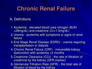
Definitions • National Kidney Foundation - Kidney Disease Outcomes Quality Initiative (NKF-K/DOQI) workgroup has defined chronic kidney disease (CKD) as the following: • The presence of markers of kidney damage for 3 months, as defined by structural or functional abnormalities of the kidney with or without decreased glomerular filtration rate (GFR), manifest by either pathological abnormalities or other markers of kidney damage, including abnormalities in the composition of blood or urine, or abnormalities in imaging tests OR • The presence of GFR <60 mL/min/1.73 m2 for 3 months, with or without other signs of kidney damage as described above.
Classifications According to a National Health and Nutrition Examination Survey (NHANES): • Stage 1 disease is defined by a normal GFR (greater than 90 mL/min per 1.73 m2) and persistent albuminuria • 2.8 % of the total United States population • Stage 2 disease is a GFR between 60 to 89 mL/min per 1.73 m2 and persistent albuminuria • 2.8 % of the U.S. population • Stage 3 disease is a GFR between 30 and 59 mL/min per 1.73 m2 • 3.7 % of the U.S. population • Stage 4 disease is a GFR between 15 and 29 mL/min per 1.73 m2 • 0.13 % of the U.S. population • Stage 5 disease is a GFR of less than 15 mL/min per 1.73 m2 or end-stage renal disease • Approximately 0.2 % of the U.S. population • Increasing at a faster rate than other stages!
Epidemiology • There are important racial/ethnic differences in the incidence and prevalence of CKD in the U.S. • 256 per million population in Caucasians • 982 per million in African Americans • 344 per million in Asian Americans and native Hawaiians and other Pacific Islanders • 514 per million in American Indians and Alaska Natives • African Americans have a disproportionately higher incidence rate of ESRD due to diabetes and glomerulonephritis compared to Caucasians. • African Americans and Hispanics tend to reach ESRD at a younger age than Caucasians (mean age 57 and 58 years, compared to 63 years).
Causes/Risk Factors Etiologies: • Diabetes - type 1 and 2 • Glomerular disease - Autoimmune diseases, systemic infections, drugs, neoplasia • Vascular disease - Hypertension, large vessel disease, microangiopathy • Tubulointerstitial disease - UTI, stones, obstruction • Cystic kidneys - Polycystic Kidney Disease
Causes/Risk Factors Risk factors for development of CKD: • Diabetes Mellitus • Hypertension • Cardiovascular disease • Family History of ESRD - even if you control for other risk factors • PCKD • Low Birth Weight - possible risk factor • Age
Causes/Risk Factors Diabetic Nephropathy: • Most common etiology of CKD • Accounts for around 50% of ESRD • Studies in type I patients indicate between 25-45% of patients develop ESRD • Pathology • Mesangial expansion • GBM thickening • Glomerular sclerosis
Causes/Risk Factors Hypertension: • Second most common cause of ESRD • Accounts for estimated 23% of ESRD between 1996-2000 • Pathology-progressive glomerulosclerosis and subsequent worsening of renal function • MRFIT trial shows odds ratio for development of ESRD to normotensives • Mild HTN=3.1, Mod=6.2, severe==11
Causes/Risk Factors Risk factors for progression of CKD to ESRD: • Hyperlipidemia - meta-analysis showed a slight decrease in GFR with lipid lowering agent • Low HDL • HTN • High protein diet - Protein restriction studies show about 0.5 yearly difference in GFR compared to control • Increased proteinuria • Obesity - observational data • African-Americans - slightly less stage I and II CKD, but greatly increased ESRD up to 5X • Poverty • Location - ESRD more common in Southern states • Male sex
Natural History • Initial insult patient-dependent - lupus, HTN, etc. • Kidney responds by adaptive hyperfiltration of undamaged nephrons - beneficial • Adaptive hyperfiltration - eventually leads to damage of these nephrons • Proteinuria results with subsequent decrease GFR • Usually asymptomatic at start of process
Natural History Progression: • NHANES data show different rates of progression to renal replacement therapy based on stage of disease • In five years • 1.1 % of stage II will progress to renal replacement • 1.3 % of stage III will progress to RR • 19.9% of stage IV will progress to RR
Complications • Hypertension • Anemia - decreased EPO production • Volume overload • Metabolic abnormalities – Acidosis, hyperkalemia, hyperphosphatemia • Renal Osteodystrophy • Dyslipidemia • Malnutrition
Screening • CKD is worth screening for because it can be detected early, and if identified, can be treated to reduce progression to ESRD – however, because CKD is relatively rare, screening of the general population is not currently recommended. • NKF-K/DOQI guidelines for CKD recommend that all individuals should be assessed, as part of routine health examinations, to determine whether they are at increased risk for developing CKD.
Screening • Screening for CKD can be simply done in higher-risk patients with a: • a urinalysis • a "spot" urine sample for protein (or if the pt is felt to be of particularly high risk, albumin) and creatinine assessment, and • a serum creatinine level. • See the K/DOQI clinical practice guidelines for chronic kidney disease for screening recommendations in various subgroups. [6]
Diagnosis - General Diagnosis In general: • Serum creatinine to estimate GFR • Protein:Creatinine ratio or Albumin:Creatinine ratio in a first morning urine specimen • Examination of urine sediment or urine dipstick for red and white blood cells • Imaging
Diagnosis Protein Quantification: • Spot Protein:Creatinine ratio – representative of daily protein excretion in grams. Ex. 240mg protein and 82mg creatinine = 2.9g protein/day. • May use albumin rather than creatinine provided proteinuria is primarily albumin • First morning collection preferred as it best represents 24 hours protein excretion • 24 hour protein collection (gold standard) • Hard for ambulatory patients • Creatinine obtained as well to assess for adequacy of collection
Diagnosis Urinalysis: • Cheap, Fast, Easy, and.. Fun? • Yes, it needs to be spun and yes, a centrifuge is hard to find • Casts • Red cell – glomerular disease such as DM • White cell - tubulointerstitial disease or acute pyelonephritis • Epithelial cell (muddy brown) – ATN and acute glomerulonephritis • Fatty – nephrotic syndrome • Protein – Albumin, (+) after >300-500mg/day • Bland – no casts, few cells, no protein more suggestive of interstitial or vascular disease
Diagnosis Imaging: • Renal Ultrasound – safe, easy • Exclude urinary tract obstruction • Polycystic kidney disease • Increased cortical echogenicity and decreased overall kidney size indicative of “medical renal disease” – irreversible • CT scan • Mainly to distinguish a simple cyst from neoplastic lesion • Renal stones (non-contrast) • MRI • Renovascular dz, RCC, contrast contraindicated
Diagnosis Renal Biopsy: • Indications vary: reversible vs. irreversible dz., persistent hematuria with rising creatinine, nephrotic syndrome (lupus nephritis), unknown etiology, transplanted kidneys • Check labs: coags, Hct, T+S • Avoided in severe HTN, bleeding disorders • Tissue examined under light, immunofluorescence, and electron microscopy
Management Reversible causes to rule out: • Hypovolemia • Hypotension (sepsis) • Drugs – aminoglycosides, NSAIDS, contrast • Urinary tract obstruction – prostate • Renal artery stenosis
Management Antihypertensive therapy: • <130/85 minimum goal • <125/75 in those with proteinuria>1 gm/day • ACE inhibitors most “renoprotective” - reduce intraglomerular pressure resulting in decreased protein excretion • ARBs as effective as ACE inhibitors in reducing proteinuria • In general a reduction in both blood pressure AND proteinuria results in preservation of renal function
Management Glycemic control: • Intensive insulin therapy thought to reverse the early glomerular hyperfiltration and hypertrophy that are thought to be important risk factors for glomerular injury • Delays development of microalbuminuria • Stabilize or decrease protein excretion in patient w/ microalbuminuria • May not slow the rate of renal injury once overt proteinuria has developed
Management Complications: • Anemia (erythropoeitin deficiency) • Target Hgb 11-12g/dl • Rule out absolute iron deficiency first (ferritin conc. <100ng/ml and Iron Sat <20%) • EPO 80-120U/kg SQ weekly – 2-4 doses • Metabolic acidosis • Tx. For arterial pH<7.25, serum bicarbonate<22 • NaHCO3 (0.5-1 meq/kg/day) • Avoid citrate as this may precipitate aluminum toxicity
Management Complications: • Hypocalcemia / Hyperphosphatemia • Serum calcium x phosphate product should be less than 50-55 • Phosphate levels between 2.7- 4.6 in CKD stage 3, 4 and 3.5-5.5 in ESRD • In general for phosphorous binders: • calcium acetate for low/normal calcium • Sevelamer for high/normal calcium • Use both for persistently elevated phosphate levels
Management ESRD options: • Hemodialysis – by far most common in U.S • Peritoneal dialysis • Generally for the more compliant patient • Needs to still make urine for volume maintenance • Kidney transplant • Preferred for younger patients • Living related>living unrelated>cadaveric
Management Renal replacement therapy: • Acute indications: A, E, I, O, U • Indications in CKD: (not absolute) • GFR < 10-15 ml/min • Fluid overload refractory to diuretics • Uremia (BUN>100) – N,V • Pericarditis • Poorly controlled HTN • Pericarditis • Malnutrition? • Access • AV fistula – least infectious risk / other complications • AV graft – (PTFE) – matures in weeks • Tunneled Catheter – lower flows, higher rates of infection
Self-Assessment Questions (see last slide for answers) • According to the NKF-K/DOQI workgroup, CKD is defined by a GFR of: • < 20 mL/min/1.73 m2 • < 40 mL/min/1.73 m2 • < 60 mL/min/1.73 m2 • < 80 mL/min/1.73 m2 • < 100 mL/min/1.73 m2
Self-Assessment Questions 2. Which of the following U.S. racial/ethnic groups has higher-than-average incidence/prevalence of CKD: • Caucasians • African-Americans • Asian-Americans • American Indians • Everyone except Caucasians
Self-Assessment Questions 3. Which out of the following is not a risk factor for progression of CKD • HTN • Living in New England • Hyperlipidemia • Poverty • Gender
Self-Assessment Questions 4. A patient with a GFR of 25 asks you what there approximate risk of needing dialysis in the next five years. You tell them • I have no idea, go see a real doctor • 5 % • 1 % • 20%
Self-Assessment Questions 5. Which of the following is not a potential complication of CKD? • HTN • Anemia • Hyperphosphatemia • Malnutrition • Never having to be admitted to 7800
Self-Assessment Questions 6. Patients with which of the following characteristics should be screened for CKD? • Diabetes • Hyperlipidemia • HTN • A and C • All of the above
Self-Assessment Questions 7. A pt with stage 3 CKD has a serum phosphate level of 7.0 and a calcium of 7.9 (albumin 3.7). What should be done about his Ca and PO4 levels? • Check a PTH and refer to endocrinology • Calcium acetate 3x/daily • Sevelamer 3x/daily • 970-SPIN
Self-Assessment Questions 8. A patient has CKD with a baseline Cr of 2.1, and DM2. BP for the last 2 visits have been 150s/90s, on no current antihypertensives, despite optimal diet and exercise. What would be your first-line agent? • Lisinopril • Amlodipine • Metoprolol • Clonidine • Irbesartan
Self-Assessment Questions 9. Which of the following is not a reasonable indication to begin dialysis in a pt with CKD A. A stable Cr of 3.0 in a 50 y/o pt B. Fluid overload refractory to diuretics C. Uremia causing N/V D. Pericarditis E. Refractory poorly controlled HTN
References • Annual data report: atlas of end-stage renal disease in the United States. 2002. Bethesda: National Institutes of Health, National Institute of Diabetes and pressure and end-stage renal disease in men. • Bello AK, et al. Chronic kidney disease: the global challenge. Lancet. 2005;365(9456):331-40. • Coresh J, Astor BC, Greene T, Prevalence of chronic kidney disease and decreased kidney function in the adult US population: Third National Health and Nutrition Examination Survey. Am J Kidney Dis. 2003;41:1-12. • Coresh J, Byrd-Holt, D, Astor, BC, et al. Chronic kidney disease awareness, prevalence, and trends among U.S. adults, 1999 to 2000. J Am Soc Nephrol. 2005;16:180. • Fried LF, Orchard TJ, Kasiske BL, Effect of lipid reduction on the progression of renal disease: a meta-analysis. Kidney Int. 2001;59:260-269. • K/DOQI clinical practice guidelines for chronic kidney disease: evaluation, classification, and stratification. Am J Kidney Dis. 2002;39:S1.
References • Kasiske BL, Lakatua JD, Ma JZ, A meta-analysis of the effects of dietary protein restriction on the rate of decline in renal function. Am J Kidney Dis. 1998;31:954-961. • Keith DS, et al. Longitudinal follow-up and outcomes among a population with chronic kidney disease in a large managed care organization. Arch Intern Med. 2004;164(6):659-63. • Klag MJ, Whelton PK, et al. Blood pressure and end-stage renal disease in men. N Engl J Med. 1996;334(1):13-8. • Levey AS, Eckardt KU, Tsukamoto Y, et al. Definition and classification of chronic kidney disease: A position statement from Kidney Disease: Improving Global Outcomes (KDIGO). Kidney Int. 2005;67:2089. • Seaquist ER, Goetz FC, Rich S, Familial clustering of diabetic kidney disease: evidence for genetic susceptibility to diabetic nephropathy. N Engl J Med 1989;320:1161-1165. • U.S. Renal Data System, USRDS 2004 Annual Data Report: Atlas of End-Stage Renal Disease in the United States, National Institutes of Health, National Institute of Diabetes and Digestive and Kidney Diseases, Bethesda, MD, 2004. Am J Kidney Dis. 2005;45(Suppl 1):S1.
Answers to Self-assessment Questions: • C • E • B • D • E • D (HL has not been identified as an RF for the development of CKD, though it has been for progression) • B • A (D would be “non-inferior” to A, but would be much more expensive) • A