Download
1 / 45
450 likes | 667 Vues
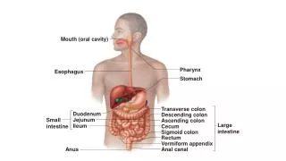
The oral cavity, encased by the lips and cheeks, plays a crucial role in digestion and speech. It is divided into two main parts: the oral vestibule and the oral cavity proper. The lips, composed of five layers including skin, muscle, and mucous membrane, form a sphincter around the mouth. The vestibule lies between the gums and teeth, while the oral cavity proper is bounded by the alveolar margins and leads to the pharynx. This guide details the structure, including the palates, mucosa, and relevant vascular connections, essential for understanding oral health and function.

E N D
The ORAL CAVITY I Dr. NabilKhouri MD, PhD
LIPS These are two fleshy folds that circumscribe the mouth and closes the cavity. At the sides they unite to form the oral commisures • 5 LAYERS • Skin– made of keratinized epith. That contains hair follicles and sebaceous glands • Superficial Fascia- contains some fats • Orbicularis Oris muscle – serves as sphincter of the mouth • Submucous tissue – contains vessels, mucous labial glands and labial branches of facial artery • Mucous membrane – innermost layer
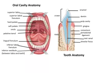
The oral cavity is conveniently divided by the arch formed by the teeth and gums into: Oral Vestibule - lies between the gums and the teeth. Oral Cavity Proper - lies behind and within the arch of teeth.
The Vestibule (Oral Vestibule) It is -Slit-like space between the cheeks and the gums It - Communicates with the exterior through the oral fissure It - Communicates with the oral cavity proper behind the 3rd molar tooth on each side only when the jaws are closed, It is - limited by the reflection of mucous membrane from lips and cheek onto the gums superiorly and inferiorly. It is – connected to the upper and lower lips by two Frenulums (upper and lower respectively) The lateral wall of the vestibule is formed by the cheek • The cheek is composed of Buccinator muscle, covered laterally by the skin & medially by the mucous membrane A small papilla on the mucosa opposite the upper 2nd molar tooth marks the opening of the duct of the parotid gland
Vestibular Boundaries Oral cavity proper • Anteriorly by the lips, • Laterally by the cheeks, • Superiorly by the Mucolabial and Mucobuccal sulcuses formed by the mucosal folds • Posteriorly and medially by the teeth and gums. Gingiva Anterior Posterior
Oral cavity proper • It is the cavity interior to the alveolar margins of the maxillae and the mandible when the mouth is closed • Anterior and lateral: It is bounded by the teeth and gums. • Posterior: Limited by the opening into the pharynx (The Oro-Pharyngeal Isthmus) • The Roof is formed by the hard palate anteriorly and the soft palate posteriorly • The Floor is formed by the mylohyoid muscle. • The anterior 2/3rd of the tongue lies on the floor.
Oral Cavity, Posterior boundaries The Oro-Pharyngeal isthmus: Is the junction of mouth and pharynx. Is bounded: By the soft palate and the palatoglossal folds superior By the dorsum of the tongue inferior
The roof of the mouth Palate • The skeleton of the hard palate • The palatine processes of the maxilla and the horizotal processes of the palatine bones. • The Greater palatine foramen carriy the greater palatine artery and nerve. • The lesser palatine foramen carriy the lesser palatine artery and nerve.
The Roof of the mouth - • Its oral surfaceiscoveredbymoucousmembranelinedbySTRATIFIED SQUAMOUS EPITHELIUM devidid in totwoparts • Anteriorlyisthehardpalatewhichformsthepartitionbetweenthe nasal and the oral cavities. • Posteriorlysoftpalatewhichisattachedtothe posterior border of thehardpalate and projectsposteriorly in tothepharynx, separatingits oral and nasal parts. • (softpalateishighlymobile and itsmovementimportant in preventingfood and drinkenteringthenasopharynx and noseduringtheact of swallowing).
The Median PALATINE RAPHE made the mucosa ends anteriorely at small elevation called THE INCISIVE PAPILLA that Overlying THE INCISSVE FOSSA (Where the nasopalatine nerve emergs in to the hard palate through this fossa)
The presence of trasverse corrugation called Maxillary Tuberosity • Located Behind the last molar tooth
STRUCTURE OF ORAL MUCOSA The oral mucosa is a Stratified Squamous Epith. • Oral epithelium Underlying connective tissue (Lamina Propria and sub mucosa) • The interface between epithelium and connective tissue is • basement membrane • This interface is irregular and is composed of downward • projections of epithelium called Rete Ridges, and upward • projection of connective tissue termed as connective tissue • Papillae
Rate Ridges Papillae
KERATINIZED AREAS • Both are distinguished from one another by color and palpation. • The hard palate is light pink while soft palate is red. • The hard palate is firm and less movable than soft palate because the mucous membrane of hard palate is tightly fixed to underlying periosteum. MASTICATORY MUCOSA 1. Hard palate 2. Gingiva
Keratinized epithelium Stratum cornium Stratum granulusum Stratum spinosum Stratum Basale
Stratum Basale --- Stratum Spinosum Single cuboidal or columnar Basal cell layer Adjacent to lamina propria The only layer where mitosis occurs Are all stem cells? Least differentiated cells Non-keratinocytes cell present • Several cells thick • Round or Ovoid cells (Prickel) • Larger and more mature than those of startumbasale • Contain - Tonofilaments - Phospholipid granules (Odland bodies) in the upper part of stratum spinosum -Increased desmosomes (shrinkage during preparation gives the spiny appearance
Stratum Granulosum --- Stratum Corneum • Cells of further increase in maturation • Cells larger and flatter • Contain - Tonofilaments & tonofibrils that occupy the cytoplasm -Keratohyline granules are present • In keratinized epithelium - Highly mature epithelial cells (squamous) -All cellular organelles and nucleus are lost (ORTHOKERATINIZATION) - In gingiva, nuclei may be retained (PARAKERATINIZATION) - cells are packed with Keratin • Keratin consist of - Tonofilaments surrounded by Filaggrin (matrix protein) • Desmosomes are weak to allow for shedding (DESQUAMATION)
In keratinized epith., as the cells of granular layer reach the junction with keratinized layer, a sudden changes occur. These changes are: 1. All the organelles with the nuclei and keratohyaline granules disappear. 2. The cells dehydrated. 3. The keratinized layer become packed with filaments, flattened, assume the form of hexagonal disks (squamous) . This pattern of maturation is termed ORTHO-KERATINIZATION. Types of keratinized epithelium
In masticatory mucosa, PARAKERATINIZATION may occur characterized with: 1. Incomplete removal of organelles from the cells of granular layer. 2. The nuclei remain (shrunken or Pyknotic). 3. Remnants of other organelles may present in the squamous cell layer.
Types of oral epithelium ORTHOKERATINISED PARAKERATINISED NON KERATINISED
KERATINOCYTES keratinocytes consists of 2 functional populations • Progenitor population – performing epithelial proliferation • Maturing population – performing epithelial maturation
Cytokeratins in oral epithelium • Keratins (previously also called cytokeratins) are filament forming proteins of epithelial cells and are essential for normal tissue structure and function • Forms the cytoskeleton of all the epithelial cells, along with microfilaments & microtubules. • Provide mechanical linkage & distribute force over wide area Based on distribution Soft keratin Hard keratin
Lines the greatest surface area of the oral mucosa, and includes the buccal and labial mucosae, the alveolar and vestibular mucosae, soft palate, tonsillar pillars, floor of the mouth, and ventral and lateral surfaces of the tongue • The Nonkeratinized squamous epithelium
Layers of Non-Keratinized Surface Epithelium • Stratum Basale • Cuboidal or columnar cells containing separate tonofilaments and other cell organelles and it is the Site of most cell divisions • Stratum Intermedium • Slightly increase in cell size as well as accumulation of glycogen in cells of the surface layer • On rare occasion, keratohyalin granules can be seen Superficial cell layer : • The cells appear more flattened. • Accumulation of glycogen. • The cells contain dispersed tonofilament. • The nuclei and some keratohyaline granules remain visible. • Diminished in number of other cell organelles. • No signs of keratinization.
Stratum corneum or superficiale- In non-keratinized epithelium - No Keratin - Tonofilaments are less and under-developed - lack keratohyline granules - this layer is less distinct • Stratum intermedium • No granular layer Superficial layer contain plump nucleus Not stain intensely with eosin
The BASEMENT MEMBRANE • Interface between connective tissue and epithelium appears thick and it includes reticular fibres. • 1-4 micrometre wide and cell free. • Ultra structurally , basement membrane is called Basal Lamina. • Basal lamina is made up of clear zone called Lamina Lucida just below the epithelial cells. • A dark zone beyond lamina lucida adjacent to the connective tissue is called Lamina Densa.
Basal Lamina : - Thick - Contains closely packed bundles of collagen fibers - Collagen fibers follow a regular course between anchoring point enabling the mucosa to resist heavy loading. • Lamina propria • Superficial papillary layer (associated with epthelial RETE RIDGES) Collagen fibers are thin and loosely arranged. • Deeper reticular layer : netlike arrangement of dense collagen fibers (nothing to do with reticulin fibers) • Papillary layer has thin and loose collagen fibers with many capillary loops • Reticular layer has collagen fibers arranged in thick bundles that are parallel to surface
The submucosa • The submucosa consists of connective tissue of varying thickness and density. It attaches the mucous membrane to the underlying structures. • Glands, blood vessels, nerves, lymph vessels and adipose tissue are present in this layer. • It is in the submucosa that larger arteries divide into smaller branches which then enter the lamina propria.
The submoucosa • Thesubmoucouslayervaries in thicknessfromoneregiontoanother and abscent in somearea. • Thisvariation in thesubmucouslayer produces 3 zone: 1. IN THE GINGIVAL REGION AND PALATINE RAPHE : • Abscent of sub-mucuoslayer, the mucosa ispink and tigthlyadherenttothebone 2. BETWEEN THE RAPHE AND THE GINGIVAL REGION ON EACH SIDE: • Isanintermediatezone in whichthesubmucosaisrelativlywelldeveloped 3. ANTERIOR TO THE INTERMEDIATE ZONE: • Thespacebetweenthe lamina propria and theperiosteumfillwithadiposetissue and themuocusmembrane. • Itisthick and pale
Specialized Lining mucosa • Covers the under surface of tongue, floor of mouth, inside of lips& cheeks, alveolar processes and soft palate. • Occupies 60% of the oral cavity • The Epithelium: - Usually thin but Thicker than that of masticatory mucosa in cheeks. - Non-keratinized in under surface of tongue, floor of mouth, cheeks, alveolar process and soft palate. - Orthokeratinized in vermilion zone and parakeratinized in intermediate zone of lips.
The floor of the mouth is a small horseshoe-shaped region situated beneath the movable part of the tongue and above the muscular diaphragm formed by the mylohyoid muscles and above this diaphragm is the genohyoid muscle.
The Floor of the Mouth Characteristics: Anterior 2/3 of the tongue, Lingual frenulum, Lingual vein, Sublingual caruncle, Sublingual folds Fimbriated fold • Covered with mucous membrane • In the midline, a mucosal fold, the frenulum, connects the tongue to the floor of the mouth • On each side of frenulum a small papilla has the opening of the duct of the submandibular gland • A rounded ridge extending backward & laterally from the papilla is produced by the sublingual gland
Inferior surface of the tongue. • The inferior surface of the tongue is covered with a thin transparent mucous membrane through which one can see the underlying veins • A sublingual caruncle (papilla) - opening of the submandibular duct 1- frenulum, 2- lingual vein, dashed-circle- sublingual gland. Arrow- Wharton's duct opening,
Lingual frenulum (The inferior surface of the tongue is connected to the floor of the mouth by a midline fold called the frenulum of the tongue. The frenulum allows the anterior part of the tongue to move freely.) Sublingual fold (passes lateraly and backwards from the papilla and overlies the sublingual gland) Papillae( openings of submandibular duct) Each sublingual compartment contains submandibular gland and dcuts, lingual and hypoglossal nerve and the siblingul vessels.
ORIGIN - Mylohyoid line of the mandible. INSERTION – Median fibrous raphe and adjacent part of hyoid body. • Action: elevates and draw forwards the hyoid bone and depress the mandible. ORIGIN - Inferior mental spines of mandible. INSERTION - Body of the hyoid bone. Mylohyoid. Geniohyoid. .
BLOOD SUPPLY • Mouth is supplied by branches from • Facial Artery • Inferior Alveolar Artery • Maxillary Artery • Infraorbital Artery • Postero superior alveolar arteries