Download
1 / 81
892 likes | 1.6k Vues
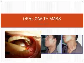
ORAL CAVITY MASS. Rivera, Laila Marie C. Rivere, Djeaune Marie Trissel B. Robosa, Dean Antonio R. Rodas, Francis Martin F. Rodriguez, Shereen Reine S. Rogelio, Ma.Graciela A. Roque, Marianne N. Ruanto, Maria Theresa R. 38 y/o M. SALIENT FEATURES. 38 y/o M 10 pack year smoking history

E N D
ORAL CAVITY MASS Rivera, Laila Marie C. Rivere, Djeaune Marie Trissel B. Robosa, Dean Antonio R. Rodas, Francis Martin F. Rodriguez, Shereen Reine S. Rogelio, Ma.Graciela A. Roque, Marianne N. Ruanto, Maria Theresa R.
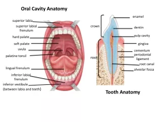
SALIENT FEATURES • 38 y/o • M • 10 pack year smoking history • (+) alcoholic beverage drinker • 2x2cm ulcer (left lower gingiva) • firm mass (left submandibular area) • mass (left lateral neck; level of the lower third of the SCM) • Oral cavity: 2x2 cm ulcer, lower gingiva near the retromolar trigone • Neck: 4x4 cm firm, well-delineated, slightly movable mass (left jugulo-digastric area); 3x3 cm firm, well-delineated, movable mass (lower third of the SCM) • Thyroid gland: (-) masses
TB adenopathy • … in developing countries… • Tuberculosis: most common cause of cervical lymph node enlargement • Peripheral lymph node tuberculosis is the most common form of extrapulmonary tuberculosis • Cervical tuberculous lymphadenopathy(scrofula) is stillthe most common cause of persistent cervical lymph node enlargement • unilateral, with little or no pain, advanced disease may suppurate and form a draining sinus • diagnosis is established by fine-needle aspiration or surgical biopsy • AFB are seen in up to 50% of cases • cultures are positive in 70 to 80% • histologic examination shows granulomatous lesions http://pmj.bmj.com/cgi/content/full/77/905/185#SEC3 http://www.javeriana.edu.co/Facultades/Medicina/pediatria/revis/ eMedicine%20-%20Tuberculosis%20%20Article%20by%20Thomas%20Herchline,%20MD.htm
Lymphoma • Lymphoma may be nodal or extranodal • A quarter of all extranodal lymphomas occur in the head and neck • Extranodal lymphoma is usually NHL • 8% of findings on supraclavicular fine-needle aspirate biopsy yield a diagnosis of lymphoma • Lymphoma is the second most common primary malignancy occurring in the head and neck • Incidence of aggressive non-Hodgkin lymphoma has risen steadily over recent decades http://emedicine.medscape.com/article/854110-overview
Non-Hodgkin’s Lymphoma • May manifest in the cervical region and lymphoid tissue of the Waldeyer ring • Appears as a mass in the oropharynx or nasopharynx • Unilateral tonsillar enlargement is highly suggestive of malignancy. • Usually arises in the tongue base • In contrast to squamous cell carcinoma, NHL is bulky, fleshy, and nonulcerating • Some patients with indolent NHLs may have large asymptomatic abdominal masses • Splenic or hepatic enlargement http://emedicine.medscape.com/article/854110-overview
Lymphadenitis from Aphthous ulcer • lymphadenitis is an infection of the lymph nodes; a complication of bacterial infection • swollen glands are usually found near the site of an underlying infection, tumor, or inflammation apthous ulcer • Apthous ulcer also known as APHTHOUS STOMATITIS • painful open sore inside the mouth, caused by a break in the mucous membrane • Etiology is unknown • Lymphadenitis may occur after skin infections or other bacterial infections, particularly those due to streptococcus or staphylococcus
Metastatic Carcinoma from oral cavity cancer • 5% percent of all cancers reported yearly • 30% of these cancers occur in the oral cavity • squamous cell carcinoma- (most common) 95% of oral cavity cancer • Risk Factors: • use of tobacco/ smoking • 80% of patients with oral SCC • risk of developing malignancy is 5-9 times greater for smokers than nonsmokers • Alcohol- 3-9 times greater risk of developing cancer • of alcohol and tobacco combined may convey a risk greater than 100 times the general population • HPV types 16 and 18 may be found in approximately 22% and 14% of oropharyngeal tumors http://www.ahns.info/patienteducation/docs/oralcavity.php http://emedicine.medscape.com/article/847678-overview
Metastatic Carcinoma from oral cavity cancer • Symptoms • most common presentation of cancer of the floor of the mouth is a painless inflamed superficial ulcer with poorly defined margin • Intermittent bleeding may occur • Advanced cases: complaints may include new or increased pain, pain on swallowing, ear pain, a change in speech, uncoordinated swallowing, or a lump in the neck • sores in the mouth, whether they are related to trauma or to a variation of canker (apthous) sores, should fully heal within three weeks http://www.ahns.info/patienteducation/docs/oralcavity.php
Metastatic Carcinoma from oral cavity cancer • Metastatic neck disease is the most important factor in the spread of head and neck squamous cell carcinoma (SCC) from primary sites • most commonly involved primary sites • larynx, oropharynx, hypopharynx, and oral cavity • Malignant tumors of the oral cavity grow rapidly, with frequent and early metastasis to the surrounding regional lymph nodes http://emedicine.medscape.com/article/850195-overview
Clinical Impression: METASTATIC CARCINOMA FROM ORAL CAVITY CANCER
What to do next • Perform a thorough head and neck exam under anesthesia • Perform triple endoscopy: (nasopharyngolaryngoscopy, bronchoscopy, esophagoscopy) • Get a biopsy of the oral cavity ulcer
Thorough head and neck exam • Biopsy of primary • Fine needle aspiration of possible neck metastasis • Imaging studies: • Chest radiograph: posteroanterior and lateral • CT/MRI of primary and neck • Panorex or dental x-ray: evaluate mandible invasion if CT/MRI not performed • Barium swallow
Thorough head and neck exam • Laboratory tests • Pre anesthesia testing • Basic liver function tests Consutations: -Radiation therapy -for adjuvant or definitive therapy considerations -Dental: pre radiation dental treatment and for post therapy
Examination under Anesthesia • Nasopharyngolaryngoscopy and pharyngoscopy • Esophagoscopy • Bronchoscopy
Nasopharyngolaryngoscopy diagnostic medical procedure that uses a flexible fibre-optic endoscope to visualize the structures inside the nasal passages, including the sinus openings, the larynx, and the vocal cords.
Pharyngoscopy • technique of placing a rigid or flexible endoscope via the mouth to visualise the pharynx (back of the throat). This technique provides direct visualisation of this structure under magnification allowing structural abnormalities to be diagnosed and any diseased areas to be accurately sampled (biopsied).
Esophagoscopy direct visual examination of the esophagus with an esophagoscope. Esophagoscopy usually is done as a diagnostic procedure for the purpose of locating and inspecting a disorder of the esophagus
Bronchoscopy • Bronchoscopy is a technique of visualizing the inside of the airways for diagnostic and therapeutic purposes. An instrument (bronchoscope) is inserted into the airways, usually through the nose or mouth, or occasionally through a tracheostomy
Findings: • Nasopharyngolaryngoscopy ⊖; Biopsy of ulcer: well-differentiated squamous cell cancer • Fine needle biopsy of neck mass: Chronic Lymphadenitis
Lymph Node Biopsy • The goal of lymphatic mapping and sentinel lymph node biopsy is to identify and remove the lymph node most likely to contain metastases in the least invasive fashion. • Sentinel node - the first node to receive drainage from the tumor site. This node is the node most likely to contain metastases, if metastases to that regional lymph node basin are present. • Recent studies evaluating treatment of an N0 neck have investigated the use of sentinel lymph node biopsy, which attempts to predict the disease status of the neck based on the first echelon of nodes that drain the tumor.
Vigilance for second primary tumors Patients diagnosed with a head and neck cancer are predisposed to the development of a second tumor within the aerodigestive tract Patients with a primary malignancy of the oral cavity or pharynx are most likely to develop a second lesion within the cervical esophagus Metastatic Work-Up
Once cancer has been proven by biopsy, a CT scan of the chest will be ordered to rule out distant metastasis • Contrast-enhanced CT and MRI of the head and neck may be performed for evaluation of the tumor and detection of occult lymphadenopathy • CT scanning - best at evaluating bony destruction • MRI - determine soft tissue involvement and is excellent at evaluating parotid and parapharyngeal space tumors • Chest radiography or chest CT is performed to rule out synchronous lung lesions • Serum tumor markers such as alkaline phosphatase and calcium may be determined, but such tests are not standard.
Positron Emission Tomography (PET) evaluates neck metastases with a sensitivity equal to that of CT • able to detect a higher percentage of lung metastases than chest radiography, bronchoscopy, or CT • but specificity ranges from 50% to 80%, and how to treat a patient with a positive PET and an otherwise negative lung workup is still in question • most common sites of distant spread are the lungs and bones, whereas hepatic and brain metastases occur less frequently • risk for distant metastases is more dependent on nodal staging than on primary tumor size
Staging • Clinical staging of the neck is based primarily on palpation, although radiographic studies, including computed tomography (CT) and magnetic resonance imaging (MRI), have been shown to be accurate in detecting positive nodes
Panoramic x-ray of the mandible scanning dental X-ray of the upper and lower jaw shows a two-dimensional view of a half-circle from ear to ear shows a patient's nasal area, sinuses, jaw joints, teeth and surrounding bone can reveal cysts, tumors, bone irregularities the mandible can also have an indentation on its lower border when the patient's masseter has been clenching and grinding shows the entire mandible, including all of its lower border Imaging for Resectability
Indications for cortical or rim resection of the mandible as determined by physical examination, CT scan, orthopantomogram, and dental films a. Tumor close to but not involving the periosteum of mandible b. Tumor involving only mandibular periosteum c. Tumor adjacent to cortical bone of mandible with no evidence of invasion beyond superficial cortex d. Tumor adjacent to dentition with no evidence of involvement of periodontal ligament
Indications forsegmental resection of the mandible as determined by physical examination, CT scan, orthopantomogram, and dental films • Invasion of the medullary space of the mandible • Tumor fixation to the occlusal surface of the mandible in the edentulous patient c. Invasion of tumor into the mandible via the mandibular or mental foramen d. Tumor fixed to the mandible following prior radiotherapy to the mandible, particularly if the tumor is located on the occlusal surface e. Tumor adjacent to carious dentition with involvement of the periodontal ligament f. Hypoplastic edentulous mandible with significant loss of vertical height precluding safe performance of a rim resection
Cortical or rim mandibulectomy – if (+) adherence to mandibular periosteum without bony erosion • Segmental resection – if (+) mandible invasion
Resection of retromolartrigone tumors: • usually requires a marginal or segmental mandibulectomy with a soft-tissue and/or osseous reconstruction in order to maximize a patient's postoperative ability for functional speech and swallowing • Ipsilateral elective and therapeutic neck dissection is performed because of the risk of metastasis to the regional lymphatics
Results of the Patient Head & neck examinations: ⊖ Chest X-ray: ⊖ Panoramic x-ray of the mandible: lytic lesion of the body of the mandible near the angle
What type of surgery is indicated? • Operative Findings: • 3 x 2 cm ulcer of the lower gingiva with invasion into the mandible • 5 X 4 cm well-encapsulated firm mass located at the submandibular triangle (level 1 to level 2) • Multiple pinkish-red, firm, grossly enlarged nodes ( 1-2 cm) along the jugular chain (levels 2 to 4) • 4 X 3 cm well encapsulated firm mass at the supraclavicular area • Operation done: • Wide excision of the ulcer with segmental mandibulectomy with modified radical neck dissection, left; the defect was reconstructed using titanium plates • Final histopath: • Well differentiated squamous cell carcinoma with metastasis to 5/20 lymph nodes, the largest measures 2 cm with extracapsular invasion; margins clear; with bony invasion
Surgical Principles • Wide resection, tumor free margins • Segmental mandibulectomy • 3 x 2 cm ulcer of the lower gingiva with invasion into the mandible • Modified radical neck dissection • Excision of lymph node levels (level I-V) with preservation of the spinal accessory nerve, internal jugular vein, and sternocleidomastoid muscle.
Modified radical neck resection • 5 X 4 cm well-encapsulated firm mass located at the submandibular triangle (level 1 to level 2) • Multiple pinkish-red, firm, grossly enlarged nodes ( 1-2 cm) along the jugular chain (levels 2 to 4) • 4 X 3 cm well encapsulated firm mass at the supraclavicular area
Modified Radical Neck Dissection • Indications: • preservation of the spinal accessory nerve (SAN), internal jugular vein (IJV), or sternocleidomastoid muscle (SCM) • N0 or N1 • N2, MRND is reasonable if any of the aforementioned nonlymphatic structures can be safely preserved.
Other Surgical Procedures • Radical Neck Dissection • Removal of all ipsilateral cervical lymph nodes (level I-V). Dissection from the inferior border of the mandible to clavicle, posteriorly to the anterior border of the trapezius muscle, and anteriorly to the lateral border of the sternohyoid muscle. Depth is to the fascia overlying the anterior scalene and levator scapulae muscles.
Indications • Multiple positive neck nodes that are clinically present in an untreated patient or in a patient treated with surgery, irradiation, chemotherapy, or a combination thereof • One or more positive neck nodes that are clinically present and extracapsular extension with involvement of the spinal accessory nerve and internal jugular vein