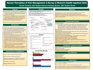
Purpose
110 likes | 423 Vues
Comparison between Dynamic contour tonometry , Goldmann applanation tonometry and Ehlers-corrected Goldmann applanation tonometry in eyes after Laser In Situ Keratomileusis Noppamas Wongpitoonpiya,MD BMA General Hospital, Bangkok, Thailand

Purpose
E N D
Presentation Transcript
Comparison between Dynamic contour tonometry, Goldmannapplanationtonometry and Ehlers-corrected Goldmannapplanationtonometry in eyes after Laser In Situ Keratomileusis Noppamas Wongpitoonpiya,MD BMA General Hospital, Bangkok, Thailand The author has no financial interest in the subject matter of this poster
Purpose • To compare intraocular pressure measurement using GoldmannApplanationTonometry (GAT) , Dynamic Contour Tonometry (DCT) and Ehlers-corrected GAT in eyes undergoing first time Laser In Situ Keratomileusis (LASIK) for correction of myopia
Methods • Central Corneal Thickness (CCT) and IOP were measured in 88 eyes of 44 patients before and 4 week after LASIK. IOP measurement was performed by GAT and DCT sequentially 3 minutes apart. • The Ehlers correction factor was applied on the GAT IOP measurement to calculate Ehlers-corrected GAT IOP.
Methods • Pearson correlation was used to evaluate the association between CCT and IOP measured by GAT and DCT.
Results • Mean preoperative IOP measured by GAT was 14.05 mmHg slightly lower compare with DCT IOP (16.27 mmHg) and Ehlers-corrected GAT (15.23 mmHg)
Results • Mean corneal ablation was 81.3 ± 23.3 micron. • After LASIK, the mean GAT measurement dropped by –4.33 mmHg ± 2.7 (SD) and the mean IOP obtained by DCT dropped by -1.17 mmHg ± 1.80 (SD). • Both GAT and DCT IOP decreased significantly after LASIK (P<0.001) but the correlation between IOP changes and CCT reduction were stronger in IOP by GAT.
Results • In contrast to IOP measured by GAT and DCT, mean Ehlers-corrected GAT was slightly higher after LASIK. • No significant correlation of CCT changes and Ehlers-corrected GAT was found in this study (P=0.90).
Conclusion • Corneal thickness alteration after LASIK affected IOP measurement by both GAT and DCT. • Ehlers-corrected GAT changes were not correlated with CCT reduction after LASIK.