BASIM ZWAIN LECTURE NOTES
Nerve. Resting membrane potential. Action potential.

BASIM ZWAIN LECTURE NOTES
E N D
Presentation Transcript
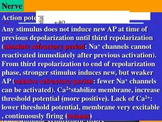
Nerve Resting membrane potential Action potential Nerve cell is neuron, 100 billion neurons ± 1006 differ in structure, chemistry & function. Neurons confer unique functions of nervous system. Glia are supporting elements, 10 times as neurons. Neurons contain cell body (soma or perikaryon) & neurites (axons & dendrites). Cell body gives rise to single axon which conducts nerve impulse from neuron to the next, up to 1 meter length, speed of impulse is a function of diameter. Dendrites: small (rarely more than 2mm)& organized symmetrically (antennae). Dendritic tree: all neurites. Neural signals: efferent (away) or afferent (towards) cell body. Classification according to conduction velocity and caliber of nerve fiber. From larger to lower caliber: Aα largest diameter & fastest conduction velocity e.g. somatic motor & proprioceptive nerve fibers Aβ e.g. sensory fibers of fine touch & fine pr. Aγ e.g. motor fibers to muscle spindle. Aδ e.g sensory fiber of acute pain, crude touch cold B e.g. preganglionic autonomic nerve fibers. C smallest diameter unmyelinated fibers e.g. sensory fibers of chronic pain, heat, gross pr. and postganglionic sympathetic fibers. Any stimulus does not induce new AP at time of previous depolarization until third repolarization (absolute refractory period: Na+ channels cannot reactivated immediately after previous activation). From third repolarization to end of repolarization phase, stronger stimulus induces new, but weaker AP (relative refractory period: fewer Na+ channels can be activated). Ca2+stabilize membrane, increase threshold potential ﴾more positive﴿. Lack of Ca2+: lower threshold potential, membrane very excitable , continuously firing ﴾tetanus﴿ Phases of AP in neurons: Depolarization phase: Sharp rise, 0 potential, overshoots,about +35 mV (activate all Na+channels, inrush of huge number of Na+, PM lose negativity Repolarization phase: Rapid fall toward negative potential (inactivate Na+ channels, continuous pumping of Na+ & passive diffusion of K+ outside) Hyperpolarization phase: Decline more negative potential than RMP,–72 mV (slow closure of K+ channels, after that, RMP again) • Ionic fluxes causing electric phenomenon of RMP: • -Na+-K+ pump: extrude 3 Na+ outside cell, intrude 2 K+ inside, much positive ions outside • Na+ channels are inactive at rest, voltage gated, activated by electric current • -Passive diffusion of K+ outside, conc. gradient, diffusion potential, major factor of RMP • -Cl- ions still inside, due to higher conc. outside • -Anionic proteins & phosphates stay inside, large size, PM impermeable Types of glia Astrocytes: Regulate EC space, remove (or restrict movement of) neurotransmitters Oligodendrocytes: Myelinating glia in CNS, form myelin sheath (wrap around axons, function in insulation), interruptions: nodes of Ranvier. Schwann Cells:PNS, myelin sheath & neurolemma Microglia:Scavenger cells get rid of foreign particle Cathodal stimuli, local responses, increase & decrease with amplitudes of stimuli, subside after removal of these stimuli. Sufficient stimulus raises the membrane potential 15 mV above RMP (i.e. –55 mV); AP phases will start & does not stop until complete cycle occurs, all-or-none rule. Membrane potential at which AP starts is threshold or firing potential. Subthreshold stimuli no or local effects, supramaximal stimuli the same effects as threshold Nerve and muscle cells are excitable cells, have the ability to reverse the negativity of their membrane potential in response to a sufficient external stimulus. The external stimulus may be electrical, chemical, physical or other types of stimuli. This change in membrane potential is action potential. Response in nerve cell is transmission of nerve impulse while response in muscle cell is contraction All living cells (animal or plant) exhibit potential difference across their plasma membranes when microelectrodes are inserted into cells, membrane interior is negative in relation to its exterior. This is resting membrane potential (RMP), due to uneven distribution of ions in & outside membrane. Classification of nerves Physioanatomic classification: afferent (sensory) & efferent (motor), subdivided into somatic & visceral neurons, general & special neurons. Accordingly, RMP of nerve cell –70 mV, skeletal muscle s –90 mV, cardiac muscle –85 mV & smooth muscle is variable but nearly –50 mV. Home Exit BASIM ZWAIN LECTURE NOTES
Nerve Propagation of action potential Synaptic and junctional transmission 12- Occlusion means that the sum of activities of several neurons working together is less than the sum of their activities when they work separately. 9- Several presynaptic neurons may converge on single postsynaptic neuron. 10-Single presynaptic neuron may diverge by its axon and its collaterals into several postsynaptic neurons. 11-Axon collaterals from postsynaptic neurons may reverberate to the presynaptic neuron(s). Acetylcholine: In neuromuscular junction (NMJ), preganglionic ANS, postganglionic parasym., basal forebrain & brain stem complexes. Synthesized from acetylcoenzyme-A & choline, catalyzed by cholineacetyltransferase enzyme (CAT), Ach is degraded in cleft by acetylcholinesterase enzyme (ACE), its receptors are nicotinic(complex of 5 αor β subunits) or muscarinic receptors. Nicotinic: in NMJ, sym. ganglia, many parts of CNS, Muscarinic in smooth muscles & glands. 4-Synaptic potentials are either excitatory (EPSP) or inhibitory (IPSP). EPSP: depolarization: cations in or anions out, while IPSP: hyperpolarization: 5-EPSP & IPSP present simultaneously in cleft. 6-Synaptic potentials are not all-or-none potentials. 7- 0.5 ms is synaptic delay 8- Spatiotemporal summation, additive or subtract-ive, numerous synaptic knobs on same neuron at same time: spatial summation, successive impulses at same synaptic knob: temporal summation. Myelin prevents leak, nodes of Ranvier act as augmentation stations, strengthen wave of depolar-ization by triggering new AP, nodes are rich in Na+ channels, saltatory conduction, jumping from node to node, one direction from soma to axon terminal (orthodromic conduction), not antidromic: absolute refractory period, proceeds forward to resting segment not backward to refractory segment. AP triggered in axon hillock, presence of large No. of Na+ channels & transmitted along axon for same reason, huge number of positive charges in firing segment of membrane is equilibrated by adjacent segments, electrotonic flow of current, very fast type of conduction in solid wires, nerve fiber is not solid wire, leaky, surrounded by sea of electrolytes, continuous triggering of AP, cost time (0.1 ms each) , unmyelinated nerves slowest conduction velocity. Criteria for classification as a NT -Must be synthesized & stored in presyn. n. -Must be released by presyn. n. upon stimulation -Application of NT directly to target cell must produce same effects as NT release Many neurons release more than single NT. Neurons that use acetylcholine: cholinergic, catecholamines: catecholinergic, serotonine: serotonergic , amino acids: amino acidergic .... Classification of neurotransmitters A. Small molecule, rapidly acting transmitters: Class I: Acetylcholine Class II: The amines (adrenaline "epinephrine", noradrenaline "norepinephrine", dopamine, serotonine and histamine). Class III: Amino acids (γ-aminobutyric acid "GABA", glycine, glutamate and aspartate). Class IV: Nitric oxide "NO". Properties of synapses: 1-One-way conduction from pre- to post-synaptic neurons because post synaptic membrane contains no synaptic vesicles. 2-Synapse is a site of neurotransduction (from electrical to chemical signal). 3-Intercellular chemical messages converted into intracellular signal. Synapses: chemical or electrical:(very rare: e.g gap junction, current flows directly through specialized protein molecule:connexon, distance between two sides of membrane is very small: 5nM). Chemical: predominant, neurotransmitters (NT) synthesized & stored in vesicles, AP opens Ca2+ channel & Ca2+ influx, increase intracellular Ca2+ conc. attract synaptic vesicles (full of NT) to presynaptic mem., signals NT to release to synaptic cleft by exocytosis. 13- Several successive EPSP may facilitate synaptic knob to depolarize while several successive IPSP may inhibit synaptic knob. 14- Continuous recurrent weak synaptic potentials may cause habituation of postsynaptic neuron, not respond for similar future stimulations. Recurrent strong stimulations accompanied by other weak stimulations may cause sensitization Vesicles fuse with active zones of presynaptic mem. NT diffuses across synaptic cleft to bind its specific receptor on postsynaptic mem. Several types of NT available and each may have types & subtypes of receptors. Postsynaptic action depends on nature of receptor. After that, NT must be inactivated by degradation, reuptake, diffusion, or bioconversion B. Neuropeptide, slowly acting transmitters: a-Hypothalamic-releasing hormones. b-Pituitary peptides. c-Peptides that act on gut and brain. d-Peptides from other tissues. Synapse is junction between two neurons. The first is presynaptic neuron and the other is postsynaptic neuron. Between them is synaptic cleft. Home Exit BASIM ZWAIN LECTURE NOTES