
BASIM ZWAIN LECTURE NOTES
E N D
Presentation Transcript
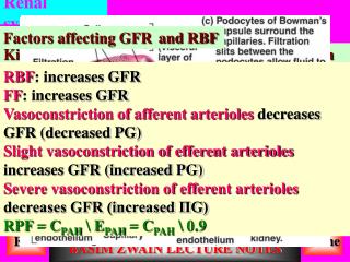
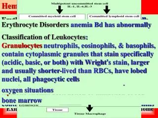
Hematology Erythropoiesis hemocytoblast, to committed cell: proerythroblast, to early erythroblasts in 3 phases: Phase 1 ribosome synthesis in early erythroblasts, Phase 2 Hb accumulation in late erythroblasts & normoblasts, Phase 3 ejection of nucleus from nor-moblasts: formation of reticulocytes, then mature erythrocytes. Circulating RBC number remains constant: balance of production & destruction. Too few RBC: tissue hypoxia, too many RBC: undesirable viscosity. Erythropoiesis is hormonally controlled, depends supplies of iron, aa, B vitamins Erythrocytes (RBCs) Biconcave discs, anucleate, essentially no organelles, filled with hemoglobin (Hb): protein that functions in gas transport, contain PM protein spectrin that: gives RBC their flexibility, allows to change shape as necessary, RBC are example of complementarity of structure & function: biconcave shape has huge surface area to vol. ratio, discounting water content, RBC are 97% Hb, ATP is generated anaerobically, so RBC do not consume O2 Functions of Blood Substance distribution (Blood transport O2, nutrients, metabolic waste, hormones ), Regulation of Bd levels of particular substances (maintains appropriate temp. by absorbing & distributing heat, normal pH in body tissues: buffer systems, adequate fluid volume), Body protection (prevents blood loss: activating plasma proteins & platelets, clot formation, blood prevents infection: synthesizing & utilizing antibodies, activating com-plement proteins, activating WBC against invader) Erythrocyte Disorders anemia Bd has abnormally low O2-carrying capacity, it is a symptom rather than a disease itself, Bd O2 levels cannot support normal metabolism, signs/symptoms: fatigue, paleness, shortness of breath & chills Anemia: Insufficient Erythrocytes hemorrhagic anemia result of acute or chronic loss of Bd, hemolytic anemia prematurely ruptured RBC Aplastic anemia destruction or inhibition of red bone marrow Erythropoiesis: Nutrient Requirements Erythropoiesis requires: Proteins, lipids, and carbohydrates, iron, vitamin B12, and folic acid The body stores iron in Hb (65%), the liver, spleen, and bone marrow, intracellular iron is stored in protein-iron complexes as ferritin & hemosiderin, circulating iron is loosely bound to the transport protein transferrin Fate and Destruction of Erythrocytes life span of RBC is 100–120 days, old RBC rigid and fragile, their Hb degenerate, engulfed by macrophages, heme & globin separated, iron is salvaged for reuse Fate of Hemoglobin heme degraded to a yellow pigment bilirubin, liver secretes bilirubin as bile to intestines, metabolized into urobilinogen, leaves body in feces: pigment called stercobilin, globin is metabolized into aa & released into circulation Anemia: Decreased Hemoglobin Content iron-deficiency anemia results from: secondary result of hemorrhagic anemia, inadequate intake of iron-containing foods, impaired iron absorption, pernicious anemia results from: deficiency of vitamin B12, often caused by lack of intrinsic factor needed for absorption of B12 Blood Plasma contains over 100 solutes, including: Proteins (albumin, globulins, clotting proteins, ...), nonprotein nitrogenous substances (lactic acid, urea, creatinine), organic nutrients (glucose, carbo-hydrates, amino acids), electrolytes (sodium, potassium, calcium, chloride, bicarbonate), respira-tory gases (oxygen and carbon dioxide) Classification of Leukocytes: Granulocytes neutrophils, eosinophils, & basophils, contain cytoplasmic granules that stain specifically (acidic, basic, or both) with Wright’s stain, larger and usually shorter-lived than RBCs, have lobed nuclei, all phagocytic cells Physical Characteristics and Volume Blood is a sticky, opaque fluid with a metallic taste color varies from scarlet (oxygen-rich) to dark red (oxygen-poor), pH of blood is 7.35–7.45, temp. is 38°C, slightly higher than “normal” body temp. it is 8% of body weight, average vol. of Bd is 5–6 L for males, and 4–5 L for females Formed Elements: RBC, WBC & platelets: Only WBCs are complete cells, RBCs have no nuclei or organelles, platelets are just cell fragments, most formed elements survive in bloodstream for only few days, most blood cells do not divide but are renewed by cells in bone marrow Leukocytes (WBCs) complete cells: less numerous than RBCs, make up 1% of the total blood volume can leave capillaries via diapedesis, move through tissue spaces, leukocytosis WBC count over 11,000 per cubic millimeter, normal response to bacterial or viral invasion Erythrocyte Function respiratory gas transport: Hb reversibly binds with O2,Hb is composed of: protein globin: 2 alpha & 2 beta chains, each bound to a heme group, each heme group bears an atom of iron, which can bind one to O2 molecule, each Hb molecule can transport 4 molecules of O2 Anemia: Abnormal Hemoglobin thalassemias absent or faulty globin chain in Hb, RBC thin, delicate, and deficient in Hb, sickle-cell anemia results from defective gene coding for an abnormal Hb called HbS: has single aa substitution in beta chain, causes RBCs to become sickle-shaped in low oxygen situations Hormonal Control of Erythropoiesis erythropoietin (EPO) release by kidneys is triggered by: Hypoxia due to decreased RBCs, decreased O2 availability, increased tissue demand for O2 , enhanced erythropoiesis increases: RBC count in circulating blood, O2 carrying ability of the blood increases Polycythemia excess RBCs increase Bd viscosity, 3 main polycythemias are: Polycythemia vera Secondary polycythemia Blood doping Hemoglobin (Hb) oxyhemoglobin Hb bound to O2, O2 loading takes place in lungs, deoxyhemoglobin Hb after O2 diffuses into tissues (reduced Hb), Carbaminohemoglobin Hb bound to CO2, CO2 loading takes place in tissues Composition of Blood: the body’s only fluid tissue, composed of liquid plasma & formed elements, formed include: erythrocytes or red blood cells (RBCs), leukocytes or white blood cells (WBCs) & platelets. Hematocrit is % of RBCs of total Bd vol. Production of Blood Cells hematopoiesis Bd cell formation, occurs in red bone marrow of axial skeleton & girdles, epiphyses of humerus & femur, hemocytoblasts give rise to all formed elements Home Exit BASIM ZWAIN LECTURE NOTES
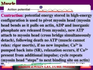
Hematology Inhibition of Clotting Factors • Fibrin acts as an anticoagulant by binding thrombin and preventing its: • Positive feedback effects of coagulation • Ability to speed up the production of prothrombin activator via factor V • Acceleration of the intrinsic pathway by activating platelets • Thrombin not absorbed to fibrin is inactivated by antithrombin III • Heparin, another anticoagulant, also inhibits thrombin activity Coagulation Phase 2: Prothr. activator catalyzes transformation of prothr. to active enz. thrombin Coagulation Phase 3: Fibrin Mesh • Thrombin catalyzes polymerization of fibrinogen into fibrin • Insoluble fibrin strands form the structural basis of a clot • Fibrin causes plasma to become a gel-like trap • Fibrin in the presence of calcium ions activates factor XIII that: • Cross-links fibrin • Strengthens and stabilizes the clot Neutrophils 2 types of granules, take up both acidic & basic dyes, cytoplasm lilac colour, peroxidases, hydrolytic enz.s & defensins (antibiotic-like prns), bacterial slayers. Eosinophils 1–4% of WBCs, red-staining, bi-lobed nuclei connected via band of nuclear material, has red to crimson(acidophilic) large, coarse, lysosome-like granules, counterattack against parasitic worm, lessen allergies, phagocyto -se immune complexes. Basophils 0.5% of WBCs, U-orS-shaped nuclei with 2-3 constrictions, large purplish-black (basophilic) granules: histamine (inflammatory chemical as VD, attracts WBCs Human Blood Groups • RBC membranes have glycoprotein antigens on their external surfaces, these antigens are: unique to individual, recognized foreign if transfused to another, promoters of agglutination: agglutinogens • Presence/absence of these antigens are used to classify blood groups • Humans have 30 varieties of RBC antigens • Antigens of ABO & Rh Bd groups cause vigorous transfusion reactions when improperly transfused • Other Bd gps (M, N, Dufy, Kell, and Lewis) are mainly used for legalities Rh Blood Groups • There are eight different Rh agglutinogens, three of which (C, D, and E) are common • Presence of the Rh agglutinogens on RBCs is indicated as Rh+ • Anti-Rh antibodies are not spontaneously formed in Rh– individuals • However, if an Rh– individual receives Rh+ blood, anti-Rh antibodies form • A second expose to Rh+ blood will result in a typical transfusion reaction Detailed Reactions of Hemostasis Coagulation Phase 1: Two Pathways to Prothrombin Activator • May be initiated by either the intrinsic or extrinsic pathway • Triggered by tissue-damaging events • Involves a series of procoagulants • Each pathway cascades toward factor X • Once factor X has been activated, it complexes with calcium ions, PF3, and factor V to form prothrombin activator Hemostasis Disorders: Bleeding Disorders • Thrombocytopenia: condition where the number of circulating platelets is deficient • Patients show petechiae (small purple blotches on the skin) due to spontaneous, widespread hemorrhage • Caused by suppression or destruction of bone marrow (e.g., malignancy, radiation) • Platelet counts less than 50,000/mm3 is diagnostic for this condition • Treated with whole blood transfusions Production of Leukocytes • Leukopoiesis is hormonally stimulated by two families of cytokines (hematopoetic factors) – interleukins and colony-stimulating factors (CSFs) • Interleukins are numbered (e.g., IL-1, IL-2), CSFs are named for the WBCs they stimulate (e.g., granulocyte-CSF stimulates granulocytes) • Macrophages and T cells are the most important sources of cytokines • Many hematopoietic hormones are used clinically to stimulate bone marrow Prevention of Undesirable Clots • Substances used to prevent undesirable clots: • Aspirin: an antiprostaglandin that inhibits thromboxane A2 • Heparin: an anticoagulant used clinically for pre- and postoperative cardiac care • Warfarin: used for those prone to atrial fibrillation • Flavonoids: substances found in tea, red wine, & grape juice that have natural anticoagulant activity Platelets • Fragments of megakaryocytes with a blue-staining outer region and a purple granular center • Granules contain serotonin, Ca2+, enzymes, ADP, and platelet-derived growth factor (PDGF) • Platelets function in clotting mechanism, forming temporary plug, helps seal breaks in blood vessels Genesis of Platelets • The stem cell for platelets is the hemocytoblast • Pathway is hemocytoblast, megakaryoblast, promegakaryocyte, megakaryocyte, and platelets Lymphocytes have large, dark-purple, circular nuclei with a thin rim of blue cytoplasm, found mostly enmeshed in lymphoid tissue (some in Bd), two types: T cells (function in immune response) & B cells(give rise to plasma cell: produce antibodies) Monocytes 4–8% of leukocytes, largest leukocytes, has abundant pale-blue cytoplasms, purple staining U-or kidney-shaped nuclei, leave circulation, enter tissue, differentiate into macrophages Macrophages highly mobile & actively phagocytic, activate lymphocytes to mount immune response Hemolytic Disease of the Newborn • Rh+ antibodies of a sensitized Rh– mother cross placenta, attack & destroy RBCs of an Rh+ baby • Rh– mother become sensitized: Rh+ blood (from previous pregnancy of Rh+ baby or transfusion) causes her body to synthesis Rh+ antibodies • The drug RhoGAM can prevent the Rh– mother from becoming sensitized • Treatment of hemolytic disease of the newborn involves pre-birth transfusions and exchange transfusions after birth Transfusion Reactions • Transfusion reactions occur when mismatched blood is infused • Donor’s cells are attacked by the recipient’s plasma agglutinins causing: • Diminished oxygen-carrying capacity • Clumped cells that impede blood flow • Ruptured RBCs that release free hemoglobin into the bloodstream • Circulating hemoglobin precipitates in the kidneys and causes renal failure Platelet Plug Formation • Platelets not stick to each other or to endothelial lining of Bd vessels. Upon damage to vessel; they: • Are stimulated by thromboxane A2 • Stick to exposed collagen fi, form a platelet plug • Release serotonin & ADP to attract more platelets • Plug is limited to area of injury by PGI2 Coagulation (a set of reactions, Bd transformed from liquid to gel, intrinsic & extrinsic pathways Diagnostic Blood Tests • Laboratory examination of blood can assess an individual’s state of health • Microscopic examination: • Variations in size and shape of RBCs – predictions of anemias • Type and number of WBCs (diagnostic of various diseases) • Chemical analysis can provide a comprehensive picture of one’s general health status in relation to normal values Leukemia • Immature white blood cells are found in the bloodstream in all leukemias • Bone marrow becomes totally occupied with cancerous leukocytes • White blood cells produced, though numerous, are not functional • Death is caused by internal hemorrhage and overwhelming infections • Treatments include irradiation, antileukemic drugs, and bone marrow transplants Hemostasis Disorders: Bleeding Disorders • Inability to synthesize procoagulants by the liver results in severe bleeding disorders • Causes can range from vitamin K deficiency to hepatitis and cirrhosis • Inability to absorb fat can lead to vitamin K deficiencies as it is a fat-soluble substance and is absorbed along with fat • Liver disease can also prevent the liver from producing bile, which is required for fat and vitamin K absorption Clot Retraction and Repair • Clot retraction – stabilization of the clot by squeezing serum from the fibrin strands • Repair • Platelet-derived growth factor (PDGF) stimulates rebuilding of blood vessel wall • Fibroblasts form a connective tissue patch • Endothelial cells multiply and restore the endothelial lining Plasma Volume Expanders • When shock is imminent from low blood volume, volume must be replaced • Plasma or plasma expanders can administered: • Plasma expanders have osmotic properties that directly increase fluid volume, used when plasma is not available, as purified human serum albumin, plasminate and dextran • Isotonic saline can also be used to replace lost blood volume • Hemophilias: hereditary bleeding disorders caused by lack of clotting factors • Hemophilia A: most common type (83% of all cases) due to a deficiency of factor VIII • Hemophilia B: results of deficiency of factor IX • Hemophilia C: mild type, caused by a deficiency of factor XI • Symptoms include prolonged bleeding and painful and disabled joints • Treatment is with blood transfusions and the injection of missing factors Developmental Aspects • Before birth, blood cell formation takes place in the fetal yolk sac, liver, and spleen • By the 7th month, red bone marrow is the primary hematopoietic area • Blood cells develop from mesenchymal cells called blood islands • The fetus forms HbF, which has a higher affinity for oxygen than adult hemoglobin Factors Preventing Undesirable Clotting • Unnecessary clotting is prevented by the structural and molecular characteristics of endothelial cells lining the blood vessels • Platelet adhesion is prevented by: • The smooth endothelial lining of blood vessels • Heparin and PGI2 secreted by endothelial cells • Vitamin E quinone, a potent anticoagulant Hemostasis Disorders: Thromboembolytic • Thrombus: a clot that develops and persist in an unbroken blood vessel • Can block circulation, resulting in tissue death • Coronary thrombosis: in Bd vessel of heart • Embolus: a thrombus freely floating in Bd stream • Pulmonary emboli can impair the ability of the body to obtain oxygen • Cerebral emboli can cause strokes Blood Transfusions • Transfusions are necessary: • When substantial blood loss occurs • In certain hemostatis disorders • Whole blood transfusions are used: • When blood loss is substantial • In treating thrombocytopenia • Packed red cells (cells with plasma removed) are used to treat anemia ABO Blood Groups • The ABO blood groups consists of: • Two antigens (A and B) on surface of the RBCs • Two antibodies in the plasma (anti-A and anti-B) • Individual with ABO Bd may have various types of antigens & spontaneously preformed antibodies • Agglutinogens and their corresponding antibodies cannot mixed without serious hemolytic reactions Formation of Leukocytes • All leukocytes originate from hemocytoblasts • Hemocytoblasts to myeloid stem cells and lymphoid stem cells • Myeloid stem cells to myeloblasts or monoblasts • Lymphoid stem cells become lymphoblasts • Myeloblasts to eosinophils, neutrophils, basophils • Monoblasts to monocytes • Lymphoblasts to lymphocytes Leukocyte Disorders: Leukemias • Cancerous conditions involving white blood cells • According to abnormal white blood cells involved: •Myelocytic leukemia (involves myeloblasts) •Lymphocytic leukemia (involves lymphocytes) •Acute leukemia (involves blast-type cells and primarily affects children) •Chronic leukemia more prevalent in older people Hemostasis • Series of reactions designed to stop bleeding • Three phases occur in rapid sequence •Vascular spasms (immediate vasoconstriction in response to injury) •Platelet plug formation •Coagulation (blood clotting) Coagulation • The final thee steps of this series of reactions are: •Prothrombin activator is formed • Prothrombin is converted into thrombin • Thrombin catalyzes the joining of fibrinogen into a fibrin mesh Developmental Aspects • Age-related blood problems result from disorders of the heart, blood vessels, and the immune system • Increased leukemias are thought to be due to the waning deficiency of the immune system • Abnormal thrombus and embolus formation reflects the progress of atherosclerosis Blood Typing • Serum containing anti-A or anti-B agglutinins is added to blood, agglutination will occur between agglutinin and corresponding agglutinogens • Positive reactions indicate agglutination Agranulocytes lymphocytes and monocytes: Lack visible cytoplasmic granules, similar structurally, but functionally distinct & unrelated cell types, have spherical (lymphocytes) or kidney-shaped (monocytes) nuclei Factors Limiting Clot Growth or Formation • Two homeostatic mechanisms prevent clots from becoming large Swift removal of clotting factors Inhibition of activated clotting factors Home Exit BASIM ZWAIN LECTURE NOTES