Thyroid Cancer 2005
Thyroid Cancer 2005. Nancy Fuller, M.D. University of Wisconsin-Madison. 52 yo woman in good health; presented with back pain of a musculoskeletal nature.


Thyroid Cancer 2005
E N D
Presentation Transcript
Thyroid Cancer 2005 Nancy Fuller, M.D. University of Wisconsin-Madison
52 yo woman in good health; presented with back pain of a musculoskeletal nature. Exam of neck: palpable right sided thyroid nodule approx 2x3 cm; gland otherwise not enlarged and no other nodules or lymphadenopathy. Ultrasound: solid nodule; uptake scan: no excess uptake in nodule TFTs: normal A FNA was performed. DX: Hurthle cell neoplasia
2. 64 yo woman with hyperlipidemia; presented for a preventive health exam with no complaints. Neck exam: 4x2 cm right sided thyroid nodule, gland otherwise normal, no lymphadenopathy. Ultrasound-solid nodule, uptake scan no excess uptake in nodule. TFTs A FNA was performed. DX: Hurthle cell neoplasia
3. 28 yo woman presented after having a thyroid nodule found incidentally on a carotid ultrasound being performed as a normal control for a study. Exam: 2x2 cm right sided thyroid nodule, gland otherwise normal, no lymphadenopathy. TFTs normal Dedicated ultrasound: solid nodule; FNA performed that day because of availability of pathology support DX: Papillary thyroid carcinoma
Learning objectives: • To learn about the epidemiology, types, behaviors, treatment and prognosis of thyroid cancer. • No financial disclosures
Epidemiology • Thyroid nodules: very common • Clinically detectable thyroid carcinoma: rare: <1% of all cancers • Female to male ratio- 2.5:1 • Median age at dx: 45-50
Overall incidence is rising: • In 1935: 1.3/100,000 women, .2/100,000 men • By 1991: 5.8/100,000 women, 2.5/100,000 men • Incidence has continued to rise in past 10 years: most rapid rate of increase in all tracked cancers
Reason for rise? • Neck irradiation: used between 1910 and 1960 • Better diagnosis? BUT: only rise is in papillary type; if better diagnosis was reason, would expect rise in all types
Algorithm for the Cost-Effective Evaluation and Treatment of a Clinically Detectable Solitary Thyroid Nodule Hegedus, L. N Engl J Med 2004;351:1764-1771
Thyroid cancer: epithelial types Differentiated: Papillary: 70-75 % of all thyroid cancers Follicular: 15-25% Undifferentiated: Anaplastic: 2-5%
Thyroid cancer: non epithelial Medullary thyroid cancer • Sporadic • Familial • MEN-2A and B Others: lymphoma, mets from breast, colon, renal and melanoma
Papillary thyroid carcinoma Pathogenesis: 1. Activation of tyrosine kinase receptors by rearrangement or gene amplification • Results in a chimeric gene • Occurs either by radiation or sporadic 2. Point mutations in BRAF gene • 10X increased risk of thyroid cancer in relatives of thyroid cancer patients: suggests a genetic link
PTC Presentation: • Solitary nodule most common • Pathology: typically unencapsulated; may be cystic Papillae: 1 or 2 layers of tumor surrounding fibrovascular core Follicles and colloid are typically absent
PTC • Psommoma bodies: scarred remnants of tumor papillae that have infarcted • Present in half of papillary thyroid carcinomas
PTC Growth and behavior: minor to major • Microcarcinoma: occult papillary carcinoma, with tumor <1cm • Found in up to 50% of glands at autopsy (rarer in children) • Incidental finding of no clinical importance
PTC • Other end of spectrum: aggressive metastasis through interthyroidal lymphatic channels to form multifocal tumors • Involves regional lymph nodes • At diagnosis: clinically detectable nodes more common in children (50%) than adults • 2-10% distant mets at dx: 2/3 pulmonary, 1/4 skeletal; also brain, kidneys, liver, adrenals
PTC Prognosis • Most patients do not die of their disease • 80-95% 10 year survival rates • Patients between 20-45: best long term survival • Patients older than 45 with lymph node recurrences are most likely to die from PTC
PTC • Prognosis is poorer in patients with large tumors: one large series showed cancer related mortality of 6%/2-3.9cm, 16%/4-6.9cm and 50%7 cm and above • Several variants have a worse prognosis: tall cell variant=1% of PTC; more aggressive and invasive
Survival Rate among 1701 Patients with Papillary or Follicular Carcinoma and No Distant Metastases at the Time of Diagnosis Schlumberger, M. J. N Engl J Med 1998;338:297-306
Follicular thyroid carcinoma • Characterized by follicular differentiation and encapsulation • Invasion of the capsule and blood vessels is the main determinant between adenomas and carcinoma • 2 main forms: minimally invasive and widely invasive • Multicentricity and lymph node involvement are less frequent than in PTC
FTC • Minimally invasive FTC behaves more like PTC • Widely invasive behaves more like anaplastic thyroid carcinoma • Hurthle Cell variant:more aggressive • FTC is more likely than PTC to be nonresponsive to I 131.
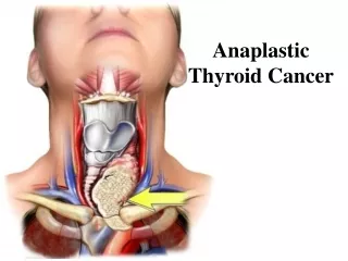
Anaplastic thyroid carcinoma • Undifferentiated tumor of thyroid follicular epithelium • Very aggressive, with a disease specific mortality approaching 100% • 2/1,000,000 annual incidence • Typical patient is older than differentiated carcinoma, mean age 65 • <10% under 50 • 60-70% women
ATC • 20% of ATC: history of differentiated thyroid carcinoma, most papillary • 10% of Hurthle cell carcinoma: has anaplastic tumor within • Up to 1/2 of ATC: history of multinodular goiter
ATC • Presentation: • Nearly all present with a thyroid mass • Regional or distant spread is present 90% of the time at dx • Lungs, bones, brain most common mets • Rapidly enlarging tumor; often causes compression symptoms like dyspnea, dysphagia, hoarseness • Constitutional symptoms like fatigue, anorexia, wt loss
ATC • 50% have palpable nodes at dx • Dx: made by FNA, then CT neck and mediastinum, CXR • Prognostic factors: tumor size <6 cm=25% 2 yr survival >6cm=3-15% 2 yr survival Others: older age, male sex, dyspnea at presentation • No effective treatment for advanced or metastatic ATC: uniformly fatal, with median survival 3-7 mo
Treatment of differentiated thyroid carcinoma • Surgery: goal is to remove all tumor tissue from neck • Total or near total thyroidectomy because of risk of multicentricity • Removal of local nodes in PTC, only palpable nodes in FTC because of lower rate of lymph node involvement
Treatment • I 131: given post op: destroys any remaining normal thyroid tissue, and may destroy occult microcarcinomas • Increases sensitivity of subsequent 1 131 total body scans • 4-6 wks after surgery a total body scan off thyroid replacement with low dose 1 131; if any uptake, a treatment dose is given (2 mCi vs. 30-100 mCi) • Radiation: only if surgical excision is impossible and tissue doesn’t take up I 131
Followup Goals of followup: • Maintain adequate thyroxine treatment • Detect persistent or recurrent cancer • Recurrences usually occur early but may occur later so follow up for life
Thyroxine treatment goals: initial serum thyrotropin level 0.1 or less, serum free T3 normal • Check U/S of thyroid area and nodal areas • Serum thyroglobulin levels: TG produced by follicular cells-should not be detectable after total ablation; presence signifies persistent or recurrent disease • 80% of patients with TG >40 have detectable foci or I 131 uptake
I 131 scanning: needs to be done after withdrawal of thyroxine tx, with TSH >30 needed • Scanning is done 3 days after I 131 given • Low risk patients with no I 131 uptake after 1 year: TSH maintained at low but detectable level (0.1-0.5)
Local or regional mets: occur in 5-20% • Excision/I 131 tx/ Radiation tx if no I 131 uptake • Distant mets: If I 131 uptake, high dose I 131 given + RT
Complications of treatment: • I 131: nausea, sialadenitis common but mild and short duration • Genetic defects: can’t be given to pregnant women • Increased risk of miscarriage in pregnancies within 1 year of tx • Overall relative risk of a second type of cancer only if high cumulative dose of I 131 and/or radiation
Medullary thyroid cancer • Much less common than epithelial thyroid cancers • Involves abnormalities of parafollicular C-cells • Most cases are sporadic
MTC • MEN 2 A: autosomal dominant disorder characterized by MTC, pheochromocytoma, and primary parathyroid hyperplasia • MEN 2 B: same inheritance; MTC + pheochromocytoma. Occurs at a younger age; more aggressive. • Familial MTC: like MEN 2 A but no other associated abnormalities
MTC • Female to male ratio=1:1 • MEN 2 A and familiar MTC: peak in index cases in 3rd decade • MEN 2 B: children and teens most common age of presentation. • Basal serum calcitonin: usually correlates with tumor mass and is almost always high with palpable tumor
MTC • MTC in MEN 2 B: more aggressive • Early onset • Surgery often not curative • Death from MTC: 50% of MEN 2 B, 10 % MEN 2 A