Download
1 / 54
540 likes | 556 Vues
This summary provides an overview of viruses and bacteria, including their structure, replication process, and viral cycles. Learn about viral nucleic acid, capsid, envelopes, and the lytic cycle.

E N D
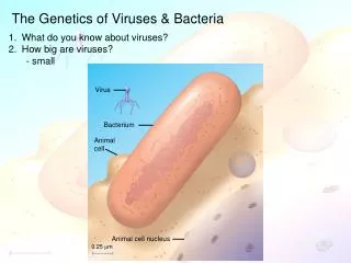
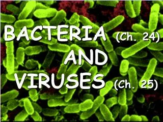
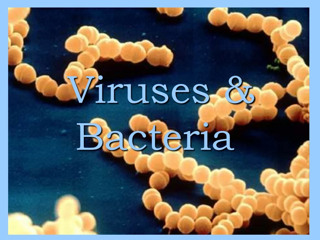
Section 18.1 Summary – pages 475-483 What is a virus? • Viruses are nonliving infectious agents composed of nucleic acids enclosed in a protein coat and are smaller than the smallest bacterium. • Most biologists consider viruses to be nonliving because they don’t exhibit all the criteria for life. • They don’t carry out respiration, grow, or develop. All viruses can do is replicate and they can’t even do that without the help of living cells. • A cell in which a virus replicates is called the host cell.
How are viruses named? • Viruses, such as rabies viruses and polioviruses, were named after the diseases they cause. • Other viruses were named for the organ or tissue they infect. Adenoidviurs infects the adenoids causing the common cold. • Today, most viruses are given a genus name ending in the word “virus” and a species name. • Sometimes code numbers are used to distinguish between similar viruses for the same host cell. • A virus that infects a bacterium is called a bacteriophage, or phage for short.
Viral Structure Section 18.1 Summary – pages 475-483 • A virus has an inner core of nucleic acid, either RNA or DNA, and an outer protein coat called a capsid. Capsid • Some relatively large viruses, such as human flu viruses, may have an additional layer, called an envelope, surrounding their capsids. • Envelopes are made from the same type of materials of the cell membrane. Nucleic acid Envelope
Section 18.1 Summary – pages 475-483 Viral Structure • Viral nucleic acid is either DNA or RNA and contains instructions for making copies of the virus. Nucleicacid • Some viruses have only four genes, while others have hundreds. Capsid • The arrangement of proteins in the capsid of a virus determines the virus’s shape as well as the type of host cell it can infect.
Viral Shape and Size Section 18.1 Summary – pages 475-483 • Polyhedral viruses resemble small crystals. • Helical or coiled springs. Capsid Nucleic acid • Virus size ranges from 20 nanometers to 250 nanometers. • Virology is the study of viruses. • Virologist is a person who studies viruses.
Section 18.1 Summary – pages 475-483 Viral Replication • Before a virus can replicate, it must enter a host cell. • A virus recognizes and attaches to a host cell when one of its proteins interlocks with a molecular shape that is the receptor site on the host cell’s plasma membrane. • In some viruses, the attachment protein is in the capsid or in the envelope. • In the T4 bacteriophage, a protein in the tail fibers recognizes and attaches the T4 to its bacterial host cell.
T4 Bacteriophage Section 18.1 Summary – pages 475-483 Capsid Nucleic acid Tail Tail fiber
Viral Replication • Each virus has a specifically shaped attachment protein. Therefore, each virus can usually attach to only a few kinds of cells. • In general, viruses are species specific, and some also are cell-type specific. For example, polio viruses normally infect only intestinal and nerve cells. • The species specific characteristic of viruses is significant for controlling the spread of viral diseases.
Viral Replication Cycles Section 18.1 Summary – pages 475-483 • Once attached to the plasma membrane of the host cell, the virus enters the cell and takes over its metabolism. Only then can the virus replicate. • Viruses have two ways of getting into host cells. • The virus may inject its nucleic acid into the host cell like a syringe injects a vaccine into your arm. The capsid of the virus stays attached to the outside of the host cell. 2. The envelope virus enters after attachment. The plasma membrane of the host cell surrounds the virus and produces a virus-filled vacuole inside the host cell’s cytoplasm. Then, the virus bursts out of the vacuole and releases its nucleic acid into the cell.
Lytic cycle Steps Section 18.1 Summary – pages 475-483 • The virus attaches to the host cell • The virus injects its nucleic acid into the host cell. • The host cell uses its own supplies to make copies of the viral nucleic acid and proteins. • New viruses are assembled and ready to be released. • The host cell breaks open and releases the new viruses to continue the cycle of infection. • The new viruses can then infect and kill other host cells. This process is called a lyticcycle. • Typical lytic viruses take about 30 minutes to complete the cycle and release 200 new viruses. • The viruses are called virulent because they cause disease immediately.
Section 18.1 Summary – pages 475-483 Lytic cycle Bacteriophage Bacterial DNA Nucleic acid Bacterial host cell B. Entry A. Attachment The bacteriophage injects its nucleic acid into the bacterial cell. E. Lysis and Release The host cell breaks open and releases new virus particles. C. Replication D. Assembly The host’s metabolic machinery makes viral nucleic acid and proteins. New virus particles are assembled.
Section 18.1 Summary – pages 475-483 Lysogenic cycle • Not all viruses kill the cells they infect. • Some viruses go through a lysogenic cycle, a replication cycle in which the virus’s nucleic acid is integrated into the host cell’s chromosome. • A lysongenic cycle begins in the same way as a lytic cycle. The virus must attach to the host cell and inject its nucleic acid. • Viral DNA that is integrated into the host cell’s chromosomes is called a provirus. • A provirus may not affect the functioning of its host cell, which continues to carry out its own metabolic activity.
Lysogenic cycle Section 18.1 Summary – pages 475-483 • However, every time the host cell reproduces, the provirus is replicated along with the host cell’s chromosome. • Therefore, every cell that originates from an infected host cell has a copy of the provirus. • The lysogenic phase can continue for many years. However, at any time, the provirus can be activated and enter a lytic cycle. Lysogenic viruses usually wait for an opportune time. A time when the body is not well rested, stressed or fatigued. • This type of virus is called temperate because it does not kill or make sick immediately.
Section 18.1 Summary – pages 475-483 Lysogenic cycle B. Provirus Formation A. Attachment and Entry Provirus Although the provirus is inactive, it replicates along with the host cell’s chromosome. C. Cell Division Bacterial host chromosome A lysogenic virus injects its nucleic acid into a bacterium. The viral nucleic acid is called a provirus when it becomes part of the host’s chromosome. LYSOGENIC CYCLE LYTIC CYCLE The provirus leaves the chromosome. The cell breaks open releasing viruses. Viral nucleic acid and proteins are made.
Disease symptoms of proviruses Section 18.1 Summary – pages 475-483 • Many disease-causing viruses have lysogenic cycles. • Three examples of these viruses are herpes simplex I, herpes simplex II that causes genital herpes, and the hepatitis B virus that causes hepatitis B. • Another lysogenic virus is the one that causes chicken pox. • Having chicken pox, gives lifelong protection from another infection by the virus. However, some of the virus may remain as proviruses in some body’s nerve cells. • Later in your life, these proviruses may enter a lytic cycle and cause a disease called shingles—a painful infection of some nerve cells.
Other diseases caused by viruses • Some other diseases caused by viruses include: • Common cold • Influenza • Measles • Mumps • Polio • Rabies Just to name a few. There are thousands of different diseases caused by viruses.
Retroviruses Section 18.1 Summary – pages 475-483 • Many viruses, such as the human immunodeficiency virus (HIV) that causes the disease AIDS, are RNA viruses—RNA being their only nucleic acid. • The RNA virus with the most complex replication cycle is the retrovirus. HIV virus
Section 18.1 Summary – pages 475-483 Retroviruses • Once inside a host cell, the retrovirus makes DNA from its RNA. • To do this, it uses reverse transcriptase, an enzyme it carries inside its capsid. • This enzyme helps produce double-stranded DNA from the viral RNA. • Then the double-stranded viral DNA is integrated into the host cell’s chromosome and becomes a provirus.
Section 18.1 Summary – pages 475-483 Retroviruses RNA Retrovirus DNA is made from the viral RNA. RNA DNA Reverse transcriptase Entering cell Provirus in host chromosome mRNA Retrovirus Cycle New virus parts Exiting cell New virus forming
Section 18.1 Summary – pages 475-483 Cancer and Viruses • Some viruses have been linked to certain cancers in humans and animals. • These viruses disrupt the normal growth and division of cells in a host, causing abnormal growth and creating tumors. • Viruses and cancer in humans: • Hepatitis B (HBV): liver infection • Human T-cell lymphotropic/leukemia virus (HTLV): certain leukemias and lymphomas • Human papillomavirus (HPV): genital warts • Epstein-Barr virus (EBV): Burkitt’s lymphoma
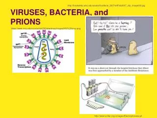
Prions and viroids Section 18.1 Summary – pages 475-483 • Researchers have recently discovered some particles that behave somewhat like viruses and cause infectious diseases. • Prions are composed of proteins but have no nucleic acid to carry genetic information. These cause abnormal clumping of proteins which results in improper functioning. • Prions are responsible for many animal diseases, such as mad cow disease BSE (Bovine Spongioform Encephalopathy) and its human equivalent, Creutzfeldt-Jakob disease.
Prions and viroids Section 18.1 Summary – pages 475-483 • Viroids are composed of a single circular strand of RNA with no protein coat. • Viroids have been shown to cause infectious diseases in several plants. • The amount of viroid RNA is much less than the amount found in viruses.
Plant viruses • The first virus to be identified was a plant virus, called tobacco mosaic virus, that causes disease in tobacco plants. • Viruses cause as many as 1000 plant diseases and are named according to their host plant. • Viruses can cause stunted growth and yield losses in their host plants. • They do not undergo lytic or lysogenic phases. • Not all viral plant diseases are fatal or even harmful. • Some mosaic viruses cause striking patterns of color in the flowers of plants.
Prevention & Treatment • Vaccinations - made from dead or inactive viruses. • Inactivated - do not replicate in the host • Attenuated - viruses that have been genetically altered so they are not capable of causing the disease. • Boosters - an additional dose of a vaccine to extend protection. Poliovirus
Prevention & Treatment • Anti-viral drugs - drugs that interfere with the viral nucleic acid synthesis. These are given to infected patients. • Best way to control disease is through prevention. • ANTIBIOTICS are NOT useful against viruses. Photo Courtesy of CDC3
Where is the Centers for Disease Control and Prevention located? Atlanta, Georgia Moscow, Russia
World Health Organization • AKA – W.H.O. • Decided in 1967 to eradicate small pox the disease from the general population. • This was officially accomplished in 1980, with the last known case in Somalia, 1977.
Emerging Viruses • These are newly discovered viruses that are emerging in different parts of the world. • They exist is isolated habitats and infect humans when those habitats are developed. • Some examples include: Ebola, HIV, and West Nile.
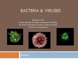
Bacteria are prokaryotic cells Bacterial structure • Kingdom Monera: contains oldest and smallest of living cells • Single-celled prokaryotes: no true nucleus or membrane-bound organelles • Ubiquitous- “They’re everywhere!” • May be harmless or pathogenic • Many species are useful industrially, ecologically, medically
Bacteria areprokaryotic cells Bacterial diversity and classification Classification: Kingdom Monera has two subkingdoms • Archaebacteria: unusual structures/habitats • Eubacteria: grouped by cell wall structure, mode of nutrition and metabolic processes
Bacteria are prokaryotic cells • Kingdom Aracheabacteria • Methanogens- unusual way to capture H2 & CO2 & convert it to CH4. Live only in anaerobic environments • Extreme Halophiles – salt loving, live in areas of high salt contents. Dead Sea & Great Salt Lake • Thermoacidophiles – acidic & extreme temperature enviroments. Volcano vents
Bacteria are prokaryotic cells • Kingdom Eubacteria • Gram stain pattern (+ or - due to cell wall) • Gram Positive – if the cell wall retains the stain & appears purple then it’s positive. Uses Crystal Violet • Gram Negative – if the cell wall does not stain & appears pink then it’s negative. Uses safranin. • Autotrophic or heterotrophic: means of obtaining nutrition (energy)
What do bacteria look like? • There are thousands of species of bacteria, but all of them are basically one of three different shapes. • Rod-shaped called bacilli. • Sphere-shaped like little balls are called cocci. • Spiral-shaped called spirilla.
Identifying bacteria Section 18.2 Summary – pages 484-495 • Diplo–is a prefix that refers to a paired arrangement of cell growth. • The prefix staphylo–describes an arrangement of cells that resemble grapes. • Strepto–is a prefix that refers to an arrangement of chains of cells.
Phylum of Bacteria • Phylum Cyanobacteria • Photosynthetic- using chemicals to capture sunlight • Once known as blue-green algae • No nucleus or chloroplasts • Too many of certain types cause eutrophication which are population blooms. This cause oxygen to be used up in bodies of water.
Phylum of Bacteria • Phylum Spirochetes • Gram negative, spiral shaped, heterotrophic • Aerobic or anaerobic • Move with a corkscrew motion • Live either freely, symbiotically or parasitically • Ex. Treponemapallidum causes syphillis
Phylum of Bacteria • Phylum Gram-Positive • All but a few of these stain positive • Examples include: Streptococci which causes strep throat and Lactobacilli which helps make yogurt. • Actinomycetes are Gram-Positive form that lives in soil and help produce antibiotics.
Phylum of Bacteria • Phylum Proteobacteria • Largest most diverse group • Three subdivisions • Enteric bacteria- Gram-negative, heterotrophic, aerobic or anaerobic environments. Examples include: Esherichia coli (E. coli) and Salmonella • Chemoautotrophs – Gram-negative that can extract energy from minerals. • Nitrogen-fixing bacteria – Gram-negative, living freely or symbiotically. Example: Rhizobium
Structure of Bacteria • Three Main Parts • Cell Wall-both types of bacteria w/a few exceptions. • Cell Membrane- used to carry out most processes due to no mitochondria • Cytoplasm-no membrane bound organelles • Some have outer coverings called capsules and pili that help adhere to the host.
Section 18.2 Summary – pages 484-495 A Typical Bacterial Cell • A typical bacterium, such as Escherichia coli would have some or all of the structures shown in this diagram of a bacterial cell. Cell Wall Capsule Chromosome Plasmamembrane Flagellum Plasmid Pilus
How do bacteria move? • Some bacteria move about their environment by means of long, whip-like structures called flagella. They rotate their flagella like tiny outboard motors to propel themselves through liquid environments. • Others may use slime, wave-like contractions or corkscrew motions to aid in movment.
What do bacteria eat? • Some bacteria are photosynthetic—they can make their own food from sunlight, just like plants. Also like plants, they give off oxygen. Other bacteria absorb food from the material they live on or in. Some of these bacteria can live off unusual "foods" such as iron or sulfur. The microbes that live in your gut absorb nutrients from the digested food you've eaten.
Bacterial Nutrition & Growth • Saprophytes-feed on dead/decaying matter. • Photoautotrophs- • Autotrophs–use sunlight • Chemotrophs- use chemicals • Obligate Anaerobes – cannot survive in O2 • Obligate Aerobes–cannot survive w/out O2 • Facultatvie Anaerobes – can survive with or without O2. • Thermophilic – bacteria that grow best b/t 40°C & 110°C. • Most bacteria prefer acidic environments of 6.0 or lower.
Bacterial reproduction Bacterial chromosome: circular, double-stranded DNA attached to cell membrane; no histones • Growth period followed by DNA replication precedes cell division. • Generation time: 20 minutes-3 hours depends on temperature, food supply, etc. • Bacteria reproduce asexually by a process known as binary fission.
Bacterial Reproduction • Three types of “sexual” Bacteria Reproduction • Transformation – bacteria takes DNA from the environment. • Conjugation – two living bacteria bind together & transfer the information to each other. • Transduction – a virus obtains a fragment of DNA from a host bacterium.
A survival mechanism Section 18.2 Summary – pages 484-495 • Some bacteria, when faced with unfavorable environmental conditions, produce endospores. • An endospore is a tiny structure that contains a bacterium’s DNA and a small amount of its cytoplasm, encased by a tough outer covering that resists drying out, temperature extremes, and harsh chemicals. • As an endospore, the bacterium rests and does not reproduce. When the time and conditions are right the endospore will begin to grow and reproduce again • Endospores can survive a temperature of 100˚C, which is the boiling point of water.
A survival mechanism Section 18.2 Summary – pages 484-495 • To kill endospores, items must be sterilized—heated under high pressure in either a pressure cooker or an autoclave. • Canned food must be sterilized and acidified. • This is because the endospores of the bacterium called Clostridium botulinum easily get into foods being canned. • If the endospores of C. botulinum get into improperly sterilized canned food, they germinate. • Bacteria grow in the anaerobic environment of the can and produce a powerful deadly poison, called a toxin, as they grow. • This deadly toxin saturates the food and, if eaten, causes the disease called botulism.
Bacteria are prokaryotic cells Bacteria as disease producers Heterotrophic bacteria can be pathogenic: • Most infect animals (some infect plants) • Use host for food; may produce toxins and/or enzyme to digest tissues of host • Transmission: bacteria enter body via food, fingers, feces, aerosols • Defense: specific (immune system) and non-specific (skin, inflammation) mechanisms