HIS Bundle ECG: Procedure, Waves, Intervals, and More
180 likes | 254 Vues
Learn about HIS bundle ECG, including definition, procedure, waves (A wave, H wave, V wave), intervals (PA interval, AH interval, HV interval), and common heart blocks and arrhythmias. Explore details on Hexaxial reference system, electrical axis of the heart, and myocardial infarction types.

HIS Bundle ECG: Procedure, Waves, Intervals, and More
E N D
Presentation Transcript
HIS Bundle ECG (HBE) • Definition – • Procedure – • Waves – A wave H wave V wave • Intervals – PA interval AH interval HV interval
R P T Q S A H Contd: PA AH HV HIS bundle ECG V
Standard lead triaxial reference system: I I III II III II 60
Unipolar lead triaxial reference system: aVL aVL aVR aVR I III II aVF aVF 60
F II III aVL aVR R 90 60 120 I 30 I 150 0 L 180 150 30 III II aVF 120 60 90 Hexaxial reference(Superimposed triaxial)system:
15 10 5 + 14 mm 0 15 10 mm 5 + 10 mm 0 -5 ELECTRICAL AXIS OF HEART: + 14 Lead I - 0 mm Lead III + 14 - 04
III I I 550 III Contd: 14 mm 00 10 mm
Heart blocks: 1st degree PR-0.38s P P 2nd degree(2:1) P P P P Missed ‘V’ beat 2nddegree(Wenkebachphenomenon)
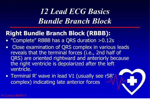
Contd: Left bundle branch block: QRS – prolonged & deformed
QRS QRS QRS Contd: Complete Heart block: Atrial rate: 107/min Ventricular rate:43/min P P P P P P P P P
Reentry Normal Circus movement Block (transient)
Arrhythmias: ‘A’ extrasystole ‘A’ tachycardia ‘A’ flutter (200-350/min) ‘A’ fibrillation (300-500/min)
Normal Wolf-Parkinson-White syndrome Lown-Ganong-Levine syndrome
‘V’ tachycardia ‘V’ fibrillation
Myocardial infarction: 1) Transmural – a) ST elevation – I, aVL, V3 to V6 b) Q wave –I, aVL & V5 to V6 c) T wave inversion 2) Subendocardial – ST depression – II, III & aVF