Download
1 / 29
290 likes | 400 Vues
This document serves as an overview of the September 28, 2011 PMT meeting, addressing key issues from previous TC discussions. It highlights demographic features, updates on imaging protocols, and the Delphi method results used in assessing hippocampal atrophy related to Alzheimer's disease. The document provides detailed metrics from scans conducted on different MRI systems, emphasizes inter-rater reliability, and data analyses highlighting the effectiveness of harmonized approaches in evaluating subjects. Key findings support the need for standardized protocols in future studies.

E N D
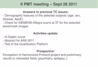
II PMT meeting – Sept 28 2011 Answers to previous TC issues: - Demographic features of the selected subjects (age, sex, disease, ApoE) - Check for SIEMENS-Allegra scans at 3T for the selected benchmark images Activities update: - III Delphi round - Abstract for AAN 2011 - Test of the Qualification Platform Prospective: Divulgation of Harmonized Protocol project and preliminary results to interested fields (psychiatry, epilepsy..).
VARIABILITY EVALUATION GOLD STANDARD 5 expert tracers 20 naive tracers Local Protocol: Experimental set (1.5T ADNI): 2 x each of the 5 Scheltens’s atrophy score x 2 sides (SAME on 3T ADNI scans) (total for each rater: 40 hippos) Benchmark Harmonized hippos: 1.5T ADNI scans 2 x each of the 5 Scheltens’s atrophy score x 2 sides (SAME on 3T ADNI scans) (total for each rater: 40 hippos) Qualification (10 tracers each SU; 10 whole hippo) Harmonized Protocol: Experimental set (1.5T ADNI): 2 x each of the 5 Scheltens’s atrophy score x 2 sides (SAME on 3T ADNI scans) (total for each rater: 40 hippos) global and local 95% confidence intervals Harmonized Protocol: 1.5T ADNI scans 2 sides x 5 Scheltens’s atrophy scores x 3 time points (0-12°month-24°month) x 3 scanners + retracing for timepoint 1 (SAME on 3T ADNI scans) (total for each rater: 240 hippos – including 40 hippos already traced) RM-ANOVA: test of main effects side, trace-retrace, atrophy, time, scanner, rater The best 5 naive tracers RM-ANOVA: test of rater and rater by center terms
Scans selection Considered as “scans”, not as “subjects”
Scans selection – Benchmark (10 sbj, 1.5T+3T= 20 scans) Considered as “subjects” Gender, Fisher’s: p=0.429; E4-carriers: p=0.283 age, ANOVA one-way: p=0.756
Scans selection – Validation (16* sbj, 1.5T + 3T, 3 time points = 90 scans) Considered as “subjects” at BASELINE *one subject: no match 1.5-3T Gender, Fisher’s: p=0.179; E4, Fisher’s: p=1 age, ANOVA one-way: p=0.641
Scans selection – Validation (16* sbj, 1.5T + 3T, 3 time points = 90 scans) Considered as “subjects” at TIME 2 *one subject: no match 1.5-3T Gender, Fisher’s: p=0.89; E4, Fisher’s: p=0.89 age, ANOVA one-way: 0.063
Scans selection – Validation (16* sbj, 1.5T + 3T, 3 time points = 90 scans) Considered as “subjects” at TIME 3 *one subject: no match 1.5-3T Gender, Fisher’s: p=0.711; E4, Fisher’s: p=0.711 age, ANOVA one-way: p=0.946
Delphi Panel – III Round I section: LANDMARKS DEFINITION
Delphi Panel – Preliminary Results from I and II rounds P=0.035 P for agreement among panelists) This preliminary model: Covers 100% of hippocampus proper Captures 100% of AD-related atrophy Has a global inter-rater of 0.94 (preliminary value across FBF-Mayo-LONI-Mainz) P=0.057 P=0.021
A model of the hippocampus, as chosen by the majority so far, but using the Horizontal criterion as first choice for the medial boundary of the hippocampal body, has the following features (right panel): Preliminary Harmonized Hippo resulting from the first two Delphi rounds but using Horizontal criterion Preliminary Harmonized Hippo resulting from the first two Delphi rounds Covers 100% of hippocampus proper Captures 100% of AD-related atrophy Preliminary inter-rater ICC (FBF-Mayo-LONI-Mainz): 0.94 Covers 99.5% of hippocampus proper Captures 99.6% of AD-related atrophy Preliminary inter-raterICC (FBF-Mayo-LONI-Mainz): 0.93
Based on this information, and on the values relating to the global preliminary models of the Harmonized Protocol, to what extent do you agree with the use of the Morphology criterion as first choice in the Harmonized Protocol? 1 2 3 4 5 6 7 8 9 Choice justification and comments:
If the agreement about the morphology criterion as first choice will be confirmed in this round, we further need to define which kind of arbitrary line should be used in a Harmonized protocol as second choice, for those images where no morphological details can be detected on the coronal plane. The possible options, as obtained from the surveyed protocols, are the oblique and the horizontal line. More exactly: The Oblique line criterion consists in continuing, using the same inclination, the oblique line delineating the boundary between the parahippocampal white matter and the hippocampal gray matter, up to the CSF of the quadrigeminal cistern, and then close the segmentation with the reset of the hippocampal tracing. The Horizontal line criterion consist in drawing a horizontal line starting from the upper point of the parahippocampal white matter, to the CSF of the quadrigeminal cistern, and then closing with reset of the hippocampal tracing. Subiculum - Oblique Subiculum - Horizontal
TAIL END Significant agreement has been achieved on the inclusion of the whole hippocampal Tail in the Harmonized Protocol, in the second Delphi round (p<0.035). Main reason for this agreement was that most caudal tissue is proper hippocampal tissue, with large contribution to global hippocampal volume and to AD related difference. (find the quantitative data about all Segmentation Unitshere).This may add sensitivity to a Harmonized Protocol. Other reasons were that caudal hippocampus can be reliably identified with 3D navigation, and that without the tail, the volume might be more dependent on individual hippocampal angulation. Some panelists note that the Crura criterion is slightly more reliable and may be easier to standardize (find all anonymous individual answers here). (You can download the presentation reporting the data from both delphi rounds and statistics here). Covers 100% of hippocampus proper Captures 100% of AD-related atrophy Preliminary inter-raterICC (FBF-Mayo-LONI-Mainz): 0.94
A model of the hippocampus, as chosen by the majority so far, but using the Crura criterion for the segmentation of the most caudal hippocampal tissue, has the following features (right panel): Preliminary Harmonized Hippo resulting from the first two Delphi rounds Preliminary Harmonized Hippo resulting from the first two Delphi rounds but using Crura criterion Covers 100% of hippocampus proper Captures 100% of AD-related atrophy Preliminary inter-raterICC (FBF-Mayo-LONI-Mainz): 0.94 Covers 88% of hippocampus proper Captures 82% of AD-related atrophy Preliminary inter-raterICC (FBF-Mayo-LONI-Mainz): 0.94
Delphi Panel – III Round II section: SEGMENTATION MODALITIES
Separation of the fimbria from the fornix “Include the white matter as fimbria as long as it runs parallel to the parahippocampal white matter located at the ventral border of the hippocampus (a, b, c), and exclude it as fornix when its inclination, in the coronal view, takes a different angle (d, e). This usually happens only in the last two slices, just caudal to the slice offering simultaneous visualization of both the inferior and superior colliculi, and before the visualization of the fornix in its full length.” a b c d e
To what extent you agree with the above definition for the segmentation of the most caudal slice that includes the fimbria in the Harmonized Protocol? If you do not agree with the above definition, please propose an alternative definition in the box below: 1 2 3 4 5 6 7 8 9 Alternative definition, or comments to the limits of the above:
Frontal view tail Medial view 3D reconstruction from Mai, J.K., Assheuer, J., Paxinos, G., 1997. Atlas of the Human Brain. Academic Press, San Diego, CA Yellow: Fasciolar gyrus Red: Andreas Retzius gyrus head tail head 3D model indicating the Andrea Retzius (AR; red) and Fasciolar gyrus (FG; yellow) within the proper hippocampal tissue. The model is built using the manual segmentation of the cytoarchitectonic boundaries of the single subject’s brain reported in the atlas by Mai JK et al., 1997 Some portions of AR and FG run internally within the hippocampal gray matter. The dorso-medial portion of the hippocampus at this level is often excluded by hippocampal segmentors, based on differences of gray matter intensity, and often independently on explicit mention of this exclusion by the protocol that is formally adopted. In our investigation, we could not find a demonstration that these different gray shades do correspond to the anatomical AR and FG. Nonetheless, when comparing the sagittal view, this tissue denoted by different gray intensity does fall in the scythe shape contiguous to the corpus callosum, that corresponds to the AR and FG as independently inferred from the Duvernoy’s atlas of the hippocampus (Duvernoy HM, 1998).
Now, different options are possible: Option n. 1) The Harmonized Protocol may include the whole vestigial tissue (Figure 1) Option n. 2) The Harmonized Protocol may exclude the vestigial tissue when, on the coronal plane, this looks different based on gray matter intensity and morphology (i.e., boundaries of regions with different gray matter intensity) (Figure 2)
Delphi III Round – Internal CSF Significant agreement (p=0.004) was achieved in the previous Delphi rounds about the fact that the CSF located internal to the hippocampus should be excluded from segmentation. Nonetheless, the criterion of connection of the CSF in the coronal plane, voted by the majority (63%), did not achieve a significant agreement. Based on the comments of panelists, the question is: To what extent do you agree with the following instruction for the Harmonized Protocol: “The CSF located internal to the hippocampus must be excluded from the segmentation when the tracer is sufficiently sure that it is CSF and not partial volume effect. This needs to be ascertained through the connection of the hypointense voxels with the CSF located external to the hippocampus, or with other hypointense voxels in rostro-caudal direction, as detected using the 3D navigation and scrolling contiguous slices. CSF spaces presumably belonging to individual differences in the morphology of the hippocampal fissure should also be excluded from segmentation” 1 2 3 4 5 6 7 8 9 Choice justification and comments:
Delphi III Round – Hardly visible structures In the previous Delphi rounds, comments of panelists were consistent with the statement that only visible tissue must be segmented in slices where structures can not be consistently seen in different raters, due to atrophy. To what extent do you agree with the following instruction for the Harmonized Protocol: “Hippocampal tissue must be segmented based on its being visible on MRI, and not based on a priori knowledge of hippocampal morphology. Nonetheless, a priori knowledge of hippocampal morphology must be used to ascertain whether hippocampal tissue can be detected in atrophic patients. For example, in some cases the subiculum is so atrophic that sometimes it can be noted only if actually searched for, adjusting image contrast and monitor settings specifically for that aim (Figure). If, after specific search guided by anatomical knowledge, thin portions of tissue can be detected, these will be segmented with a thin trace, trying not to overestimate its volume. If no tissue can be detected in such search, the segmentation should not complete the hippocampal shape based on a priori knowledge, but should only include the tissue that is actually visible after all possible checks.” 1 2 3 4 5 6 7 8 9 Choice justification and comments:
Delphi III Round – Image orientation Significant agreement (p=0.012) was achieved on the fact that the image orientation for hippocampal segmentation should be aligned parallel to the long axis of the hippocampus. Now, it should be defined whether the MRI images should be oriented: • Parallel to the long axis of one of the two hippocampi (say, the left) of each subject (same orientation for both hippocampi in each subject). • Parallel to the long axis of the currently segmented hippocampus, in each subject (two orientations used in each subject). • Along the axis representing the mean angle among the right and the left long axes of the hippocampi of each subject (same orientation for both hippocampi in each subject). * * * Choice justification and comments:
AAN 2012 “Definition of landmarks for a Harmonized Protocol for the manual segmentation of the hippocampus on MRI: preliminary results from the EADC-ADNI working group” Marina Boccardi, Martina Bocchetta, Liana Apostolova, Josephine Barnes, George Bartzokis, Gabriele Corbetta, Charles deCarli, Leyla DeToledo-Morrell, Michael Firbank, Rossana Ganzola, Lotte Gerritsen, Wouter Henneman, Ronald Killiani, Nikolai Malykhin, Patrizio Pasqualetti, Jens Pruessner, Alberto Redolfi, Nicolas Robitaille, Hilkka Soininen, Daniele Tolomeo, Lei Wang, Craig Watson, Henrike Wolf, Simon Duchesne, Clifford R. Jack Jr, Giovanni B. Frisoni.
1 tracer VARIABILITY EVALUATION GOLD STANDARD VALIDATION vs PATHOLOGY 5 expert tracers 20 naive tracers Training (tracing 20 hippos on 1.5T ADNI scans with each SU) (SAME on 3T ADNI scans) Local Protocol: Experimental set (1.5T ADNI): 2 x each of the 5 Scheltens’s atrophy score x 2 sides (SAME on 3T ADNI scans) (total for each rater: 40 hippos) Local Protocol: 1.5T 3D T1-weighted scans from (Bobinski et al., 2000) pathologically verified set (total for rater: 30 hippos) Delphi panel → harmonized prot Benchmark Harmonized hippos: 1.5T ADNI scans 2 x each of the 5 Scheltens’s atrophy score x 2 sides (SAME on 3T ADNI scans) (total for each rater: 40 hippos) Qualification (10 tracers each SU; 10 whole hippo) Qualification Harmonized Protocol: 1.5T 3D T1-weighted scans from (Bobinski et al., 2000) pathologically verified set (total for rater: 30 hippos) Harmonized Protocol: Experimental set (1.5T ADNI): 2 x each of the 5 Scheltens’s atrophy score x 2 sides (SAME on 3T ADNI scans) (total for each rater: 40 hippos) global and local 95% confidence intervals Harmonized Protocol: 1.5T ADNI scans 2 sides x 5 Scheltens’s atrophy scores x 3 time points (0-12°month-24°month) x 3 scanners + retracing for timepoint 1 (SAME on 3T ADNI scans) (total for each rater: 240 hippos – including 40 hippos already traced) RM-ANOVA: test of protocol main effect RM-ANOVA: test of main effects side, trace-retrace, atrophy, time, scanner, rater The best 5 naive tracers RM-ANOVA: test of rater and rater by center terms
Next: symposia Divulgation of Harmonized Protocol project and preliminary results to interested fields (psychiatry, epilepsy..) Society of Biological Psychiatry (SOBP) Philadelphia May 2012 – Deadline for symposium is Oct 24, 2011 (Csernansky?) Epilepsy/Neurology meeting other than AAN? Other?