The Autonomic Nervous System
690 likes | 794 Vues
The Autonomic Nervous System. Autonomic Nervous System (ANS). The ANS consists of motor neurons that: Innervate smooth and cardiac muscle and glands Make adjustments to ensure optimal support for body activities Operate via subconscious control Have viscera as most of their effectors.

The Autonomic Nervous System
E N D
Presentation Transcript
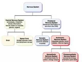
Autonomic Nervous System (ANS) • The ANS consists of motor neurons that: • Innervate smooth and cardiac muscle and glands • Make adjustments to ensure optimal support for body activities • Operate via subconscious control • Have viscera as most of their effectors
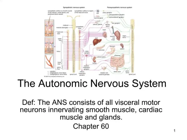
Divisions of the ANS • Sympathetic division (thoracolumbar, “fight or flight”) • Thoracic and lumbar segments • Parasympathetic division (craniosacral, “rest and repose”) • Preganglionic fibers leaving the brain and sacral segments • Enteric nervous system (ENS) • May work independently
Sympathetic and Parasympathetic • Often they have opposing effects • May work independently • May work together each one controlling one stage of the process
ANS Versus Somatic Nervous System (SNS) • The ANS differs from the SNS in the following three areas • Effectors • Efferent pathways • Target organ responses
Effectors • The effectors of the SNS are skeletal muscles • The effectors of the ANS are cardiac muscle, smooth muscle, and glands
Efferent Pathways • Heavily myelinated axons of the somatic motor neurons extend from the CNS to the effector • Axons of the ANS are a two-neuron chain • The preganglionic (first) neuron has a lightly myelinated axon • The ganglionic (second) neuron extends to an effector organ
Neurotransmitter Effects • All somatic motor neurons release Acetylcholine (ACh), which has an excitatory effect • In the ANS: • Preganglionic fibers release ACh • Postganglionic fibers release norepinephrine or ACh and the effect is either stimulatory or inhibitory • ANS effect on the target organ is dependent upon the neurotransmitter released and the receptor type of the effector
Sympathetic division anatomy • Preganglionic neurons between segments T1 and L2 – lateral gray horn of spinal cord • Preganglionic fibers • Short • Travel in the ventral root and spinal nerve • Ganglionic neurons in ganglia near vertebral column • Specialized neurons in adrenal glands • Postganglionic fibers • Long fibers
Sympathetic ganglia • Sympathetic chain ganglia (paravertebral ganglia) • Collateral ganglia (prevertebral ganglia) • Adrenal medulla
Organization and anatomy of the sympathetic division • Segments T1-L2, ventral roots give rise to myelinated white ramus • Leads to sympathetic chain ganglia
Postganglionic fibers of the sympathetic ganglia • Some fibers will return to the spinal nerve through a gray ramus and will innervate skin, blood vessels, sweat glands, adipose tissue, arrector pili muscle (body wall structures) • Postganglionic fibers coming from chain ganglia will form sympathetic nerves that will innervate thoracic organs
Collateral ganglia • Preganglionic fibers will pass through the sympathetic chain without synapsing • Preganglionic fibers will synapse within collateral ganglia • Preganglionic fibers synapsing within collateral ganglia will from Splanchnic nerves
Collateral ganglia • Celiac ganglion • Postganglionic fibers innervates stomach, liver, gall bladder, pancreas, spleen • Superior mesenteric ganglion • Postganglionic fibers innervates small intestine
Collateral ganglia • Inferior mesenteric ganglion • Postganglionic fibers innervate the large intestine • Inferior hypogastric • Postganglionic fibers innervates urinary bladder , sex organs
Adrenal medulla • Preganglionic fibers will pass through sympathetic chain ganglia and collateral ganglia without synapsing • Preganglionic fibers will then synapse on adrenal medulla • Adrenal medulla will secrete • Epinephrine • Norepinephrine
Adrenal medulla • Neurotransmitter will go into general circulation • Their effects last longer than those produced by direct sympathetic innervation
Role of the Sympathetic Division • The sympathetic division is the “fight-or-flight” system • Involves E activities – exercise, emergency • Promotes adjustments during exercise • Blood flow to organs is reduced, flow to muscles is increased
Role of the Sympathetic Division • Its activity is illustrated by a person who is threatened • Heart rate increases, and breathing is rapid and deep • The skin is cold and sweaty, and the pupils dilate
Parasympathetic division (craniosacral division) • Preganglionic neurons in the brainstem(nuclei of cranial nerves III, VII, IX, X) and sacral segments of spinal cord (S2-S4) • Ganglionic neurons in peripheral ganglia located within or near target organs • Terminal ganglion • Intramural ganglion
Organization and anatomy of the parasympathetic division • Preganglionic fibers leave the brain as cranial nerves III, VII, IX, X • Cranial nerve X provides 75% of the parasympathetic outflow • Sacral neurons form the pelvic nerves
Parasympathetic activation • Effects produced by the parasympathetic division • relaxation • food processing • energy absorption • Pupil constriction • Constriction of respiratory passageway • Decrease heart rate and blood pressure • Stimulates defecation and urination
Summary: The Anatomical Differences between the Sympathetic and Parasympathetic Divisions
Sensory Visceral Neurons • Are found in: • Sensory ganglia of cranial nerves • Dorsal root ganglia • Sympathetic ganglia • Afferent visceral fibers are found in: • Cranial nerves VII, IX, X • Autonomic nerves • Spinal nerves
Visceral Reflexes • Visceral reflexes have the same elements as somatic reflexes • They are always polysynaptic pathways
Referred Pain • Pain stimuli arising from the viscera are perceived as somatic in origin • This may be due to the fact that visceral pain afferents travel along the same pathways as somatic pain fibers
Neurotransmitters and Receptors • Acetylcholine (ACh) and norepinephrine (NE) are the two major neurotransmitters of the ANS • ACh is released by all preganglionic axons and all parasympathetic postganglionic axons • Cholinergic fibers – ACh-releasing fibers
Neurotransmitters and Receptors • Adrenergic fibers – sympathetic postganglionic axons that release NE • Neurotransmitter effects can be excitatory or inhibitory depending upon the receptor type
Neurotransmitters and parasympathetic functions • All parasympathetic fibers release ACh • Short-lived response as ACh is broken down by AChE and tissue cholinesterase • Postsynaptic membranes have two kinds of receptors: muscarinic and nicotinic
Neurotransmitters and parasympathetic functions • Muscarinic • Parasympathetic target organs • Postganglionic cholinergic fibers • Cardiac muscle • Smooth muscle • Excitatory or inhibitory effects • Depends on the receptor type of the target organ
Nicotinic Receptors • Nicotinic receptors are found on: • Surface of skeletal muscles • All ganglionic neurons of both sympathetic and parasympathetic divisions • Ganglionic neurons of the adrenal medulla • The effect of ACh binding to nicotinic receptors is always stimulatory by opening Na channels
Adrenergic Receptors • The two types of adrenergic receptors are alpha and beta • Each type has two or three subclasses (1, 2, 1, 2 , 3)
Adrenergic Receptors • Alpha 1 • Constrict blood vessels of: skin, mucosa, abdominal viscera, kidney, salivary glands, etc. • Dilates pupil • Constrict involuntary sphincters • Excitatory