Download
1 / 23
270 likes | 789 Vues
Bacteriophage Families with a detailed description of Models Phages Myoviridae – Mu. Viro102: Bacteriophages & Phage Therapy 3 Credit hours NUST Centre of Virology & Immunology. Bacteriophage Families. Myoviridae Family. Group I viruses Single molecule of ds linear DNA .

E N D
Bacteriophage Families with a detailed description of Models PhagesMyoviridae – Mu Viro102: Bacteriophages & Phage Therapy 3 Credit hours NUST Centre of Virology & Immunology
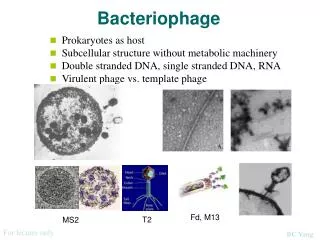
Myoviridae Family • Group I viruses • Single molecule of ds linear DNA. • Non enveloped. • diameter of about 55-110 nm. • Genome size ranges from 33.6 – 170 kb. • The genome contains unusual bases, they are 5-hydroxy-methyl cytosine (instead of cytosine). This helps in protecting the phage from the host defence system i.e. Restriction enzymes.
Mu: Discovery • Discovered in E. coliby Larry Taylor (1963). • Given the name Mu, for mutator because of its ability to cause mutations. It is known to cause mutations at high rate. • The mutations proved to be insertions to Mu at random sites in the host genome disrupting the functioning of different genes.
Mu: An Introduction • linear ds DNA genome of 40-Kb and more than 35 genes. • Capable of both Lytic & lysogenic life cycle. • Most important feature is its capability to ‘move’ within host genome, a process referred to as transposition. This phage replicates by transposition. • Mu uses multiple rounds of replicate transposition to amplify it during lytic growth. • During lytic cycle Mu completes about 100 rounds of transposition per hour, making it most efficient transposition known. • The head of Mu phage has the capability to carry 2 kb extra genome. This is because of headful packaging mechanism.
DNA Transposition Transposons are sequences of DNA that can move around to different positions within the genome of a single cell, a process called transposition. In the process, they can cause mutations & change the amount of DNA in the genome. Class I mobile genetic elements (aka retrotransposons) copy themselves by first being transcribed to RNA, then transcribed back to DNA by reverse transcriptase, & then being inserted at another position in the genome (copy paste mechanism). Class II mobile genetic elements (aka DNA transposons) move directly from one position to another using a transposase to "cut and paste" them.
Mu: Structure • Isometric, icosahedral head • a knob like neck • a contractile tail • a baseplate • six short tail fibers Figure 1. Electron micrograph of a Mu virion, negatively stained with uranyl acetate. Scale bar represents 50 nm.
Each Mu is packaged from a different site in the host genome, so the host DNA on the ends of Mu is unique in every different phage head.
Mu: Genome When Mu DNA is packaged into a phage head it includes about 50-150 bp of host DNA at the left end and a variable amount of host DNA (2kb) on the right end.
Cont’d • Gene C encodes the Repressor • Gene A encodes Transposase that is responsible for integration, replication transposition, and excision of Mu DNA • Gene B encodes enhancer of transposition. • Mom gene is responsible for protecting the virus against restriction endonucleases.
Mu: Host Recognition • The Mu genome contains a region, called G, that can invert. Its about 3000 bp reigon. • Each orientation of this DNA fragment corresponds to the synthesis of different proteins involved in the host specificity of the viral particle. • The two different kinds of particles are called • Mu G(+) • Mu G(-)
A phage specific Gin( G inversion) protien is resposible for switching, which occur time to time.
Mu: Integration into host genomeLittle is known about how this occurs apart from the fact that the bacterial sequences at either end of the Mu genome are lost in the process
Mu: Transposition Transposition requires two phage encoded proteins: Transposase (encoded by gene A) Transposition enhancer (encoded by gene B). In bacterial cells, Mu transposition can be Non Replicative: Initial insertion of the Mu genome into host chromosome (Lysogeny). Replicative: Mu phage makes copies of its own genome while inside the host chromosome (Lytic).
Mu uses target immunity to avoid transposing into its own DNA • Transposition in its own genome causes disruption in its genes. • That is solved by target immunity. • Achieved by interplay between MuA trasposase and MuB ATPase. • MuA inhibit MuB from binding to nearby DNA sites. This inhibition requires ATP hydrolysis • MuB helps MuA to find a target site for transposition • For Mu sequence within approximately 15 kb of an existing Mu insertion are immune to new insertion.
Things to remember about Mu • Mu phage can tranpose. • Mu phage genome doesnot concatamerize. • Mu phage replicates by semi conservative replication in the host genome. • Mu phage has a diverse host range because of G fragment in genome.