Download

1 / 31
310 likes | 515 Vues
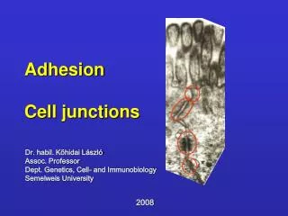
Last Class Junctions: Occluding Junctions, Anchoring Junctions, Communicating Junctions 2. Occluding Junctions: Tight Junction 3. Anchoring Junctions: adherens Junction 4. Communicating Junctions: Gap Junction 5. adherens junction, cadherins. Manipulating DNA, RNA and Proteins

E N D
Last Class • Junctions: Occluding Junctions, Anchoring Junctions, Communicating Junctions • 2. Occluding Junctions: Tight Junction • 3. Anchoring Junctions: adherens Junction • 4. Communicating Junctions: Gap Junction • 5. adherens junction, cadherins
Manipulating DNA, RNA and Proteins • Isolation of cells and cell culture • DNA manipulation • Protein measurement and analysis
Isolating Cell and Cell Culture • Tissue are disassembled by (1) preteolytic enzymes to separate cells from ECM, e.g. trypsin and collagenase, (2) together with reagents to chelate Ca2+. • 1. Centrifuge to separate based on size. • 2. adhesion strength • 3. Antibody binding • A. FACS (fluorescence activated cell sorter). • B. Microdissection (Laser capture microdissection)
FACS Machine Fluorescence cells labeled with negative charges, Non-fluorescence cells with positive charges, Clustered cells no charges due to their large sizes
Laser capture microdissection A laser beam to excise a region of interest and select for further culture
In vitro Cell Cultures Some terminologies: in vitro, in vivo, primary culture Phase contrast images of fibroblasts (A) and myoblasts (B)
In vitro Cell Cultures Phase contrast images of oligodentrocytes (C) and tobacco cells (BY2 immortal cell line) (D)
Preparation of Hybridomas that secrete desired monoclonal antibodies
Cell Fraction Centrifuge Chromatography Polyacrylamide Gel Electrophoresis Mass Spectrometry
Further separation Velocity sedimentation (size and shape) and equilibrium sedimentation (buoyant density)
Three Typical Chromatography Methods Affinity Chromatography usually gives better specificity
Protein purification with the combination of chromatography approaches
2D PAGE Coomassie blue stainging
2D Western Blot Tobacco cells, (A) gel staining (B) phospho-thereonine
Mass Spectrometry Matrix-assisted laser desorption ionization-time-of-flight spectrometry (MALDI-TOF)