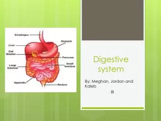
Digestive system
Presented By Logan Sumner and Tony Elias. Digestive system. Introduction. Digestion refers to the mechanical and chemical breakdown of foods so that nutrients can be absorbed by cells. The digestive system carries out the process of digestion.


Digestive system
E N D
Presentation Transcript
Presented By Logan Sumner and Tony Elias Digestive system
Introduction • Digestion refers to the mechanical and chemical breakdown of foods so that nutrients can be absorbed by cells. • The digestive system carries out the process of digestion. • The digestive system consists of the alimentary canal, leading from mouth to anus, and several accessory organs whose secretions aid the processes of digestion.
General Characteristics of the Alimentary Canal • The alimentary canal is a muscular tube that passes through the body's ventral cavity.
Structure of the Wall • The wall of the alimentary canal consists of four layers, with only slight variations according to the functions of specific sections of the canal. • The inner layer is the mucosa, protects tissues of the canal and carries on secretion and absorption. • The next layer is the submucosa it nourishes the surrounding layers of the canal. • The muscular layer consists of inner circular fibers and outer longitudinal fibers that move food through the canal. • The outer layer is composed of visceral peritoneum that protects underlying tissues and secretes serous fluid to prevent the canal from sticking to other parts in the abdominal cavity.
Movements of the Tube • The functions of the alimentary canal are to provide a mixing movements and propelling movements • Mixing movements are when smooth muscles contract rhythmically in small sections of the tube • Propelling movements are a wavelike motion called peristalsis, which is caused by contraction behind a mass of food as it relaxes it allows the mass to enter the next segment of the tube
Mouth • The mouth is the first portion of the alimentary canal; it function is to receive food and begins mechanical digestion by mastication
Cheeks and Lips • Cheeks form the lateral walls of the mouth. • The lips surround the mouth opening. • The lips have sensory receptors to help to judge the temperature and texture of food.
Tongue • The tongue is a thick, muscular organ covered by mucous membrane with taste buds within papillae; it is attached to the floor of the mouth by the frenulum. • The papillae provides friction for moving food around in the mouth. • Lingual tonsils are lymphatic tissues located at the root of the tongue.
Palate • The palate forms the roof of the oral cavity and has an anterior hard palate and posterior soft palate. • The soft palate and uvula job is to close the nasal cavity during swallowing. • The palatine tonsils are associated with the soft palate in the back of the mouth because they help to protect the body against infection.
Teeth • Teeth develop in sockets within the alveolar processes of the maxillary and mandibular bones in pairs • There are 20 primary teeth in the order they appear and are replaced by 32 secondary teeth. • Through the actions of chewing, teeth break food into smaller pieces, which begins mechanical digestion.
Teeth Cont. • Different teeth are adapted to handle food in different ways, and include incisors, cuspids, bicuspids, and molars. • Each tooth consists of a crown and a root, and is made of enamel, dentin, pulp, cementum, nerves, and blood vessels. • A tooth is held tight in its socket by a periodontal ligament.
Salivary Glands • The salivary glands secrete saliva, which moistens and dissolves food particles, binds them together, allows tasting, helps to cleanse the mouth and teeth, and begins carbohydrate digestion
Salivary Secretions • Salivary glands contain serous cells that produce a watery fluid with amylase, and mucous cells that produce lubricating and binding mucus. • Salivary glands receive parasympathetic stimulation which triggers the production of a large volume of saliva at the sight or smell of food.
Major Salivary Glands • The parotid glands, are the biggest of the major salivary glands, secretes a clear, watery fluid rich in amylase • The submandibular glands, secrete a more viscous fluid • The sublingual glands, are the smallest of the major salivary glands and secrete a thick and stringy saliva
Pharynx and Esophagus • The pharynx is a cavity behind the mouth, and the esophagus is the muscular tube leading to the stomach • The pharynx job in both the digestive and respiratory systems is the same
Structure of the Pharynx • The pharynx connects the nasal and oral cavities with the larynx and esophagus and is divided into a nasopharynx, oropharynx, and laryngopharynx
Swallowing Mechanism • Swallowing reflexes are divided into three stages. • Food is mixed with saliva and goes into the pharynx with the tongue. • Sensory receptors in the pharynx sense food, which start swallowing reflexes. • Peristalsis transports the food in the esophagus to the stomach.
Esophagus • The esophagus is a straight, collapsible passageway leading to the stomach • Mucous glands are scattered throughout the submucosa of the esophagus and produce mucus lubricate the inner lining of the tube • The lower esophageal sphincter helps prevent regurgitation of the stomach contents
Stomach • It is a J-shaped muscular organ that receives and mixes food with digestive juices, and moves food to the small intestine • It is divided into cardiac, fundic, body, and pyloric regions and a pyloric canal • Pyloric sphincter controls release of food from the stomach into the small intestine
Gastric Secretions • Gastric glands within the mucosa of the stomach open as gastric pits. • Gastric glands generally contain three types of secretory cells Mucous cells, Chief cells, Parietal cells
The Cells • Mucous cells produce mucus that protects stomach lining • Chief cells secrete pepsin as inactive pepsinogen, which is activated by contact of hydrochloric acid • Parietal cells secrete hydrochloric acid
Regulation of Stomach Juice • Gastric secretions are enhanced by parasympathetic impulses and the hormone gastrin, released from gastric glands. • As food enters the small intestine, secretion of gastric juice from the stomach wall is reflex inhibited. • Presence of fats and proteins in the upper small intestine causes the release of cholecystokinin from the intestinal wall, which decreases gastric mobility.
Gastric Absorption • The stomach absorbs small quantities of water, certain salts, alcohol, and some lipid-soluble drugs
Mixing and Emptying • Following a meal, mixing actions of the stomach turn the food into chyme and move it toward the pyloric region using peristaltic waves • The rate the stomach empties depends on the fluidity of the chyme and the type of food • After chyme fills the duodenum, stretching its walls triggers the enterogastric reflex, which inhibits peristalsis and slows the rate at which chyme enters the small intestine
Pancreas • The pancreas has an exocrine function of producing pancreatic juice that aids digestion
Structure of the Pancreas • Closely associated with the small intestine. • Cells that produce pancreatic juice, are called pancreatic acinar cells, makes up of the pancreas. • Acinar cells cluster around tiny tubes that merge to form bigger ones, and then make the pancreatic duct. • The pancreatic and bile ducts join and empty into the small intestine
Pancreatic Juice • Pancreatic juice contains enzymes that digest carbohydrates, fats, proteins, and nucleic acids. • Pancreatic enzymes include pancreatic amylase, pancreatic lipase, trypsin, chymotrypsin, carboxypeptidase, and two nucleases. • Enzymes are released inactively and are activated when they reach the small intestine.
Regulation of Pancreatic Secretion • The nervous and endocrine systems regulate release of pancreatic juice • Secretin from the duodenum stimulates the release of pancreatic juice Cholecystokinin from the wall of the small intestine stimulates the release of pancreatic juice, and digestive enzymes
Liver • The reddish-brown liver, located in the upper right quadrant of the abdominal cavity, is the body’s largest internal organ
Liver Structure • The liver is divided into right and left lobes, and is enclosed by a fibrous capsule • Each lobe is separated into hepatic lobules consisting of hepatic cells radiating from a central vein • Hepatic sinusoids separate groups of hepatic cells. • Blood from the hepatic portal vein carries blood rich in nutrients to the liver • Kupffer cells carry on phagocytosis in the liver • Secretions from hepatic cells are collected in bile canals that join to become hepatic ducts and form the common hepatic duct
Liver Functions • The liver is responsible for many metabolic activities, such as the metabolism of carbohydrates, lipids, and proteins • The liver filters the blood, removing damaged red blood cells and foreign substances, and removes toxins • The liver's job in digestion is to secrete bile
Composition of Bile • Bile is a yellowish-green liquid that hepatic cells secrete; includes water, bile salts, bile pigments, cholesterol, and electrolytes • Bile pigments are the breakdown products from red blood cells • Only bile salts have a digestive function
Gallbladder • The gallbladder is a pear-shaped sac on the interior surface of the liver • It is connected to the cystic duct, which joins the hepatic duct; which merge to form the common bile duct and leads to the duodenum • A sphincter muscle controls the release of bile from the common bile duct
Regulation of Bile Release • Bile does not normally enter the duodenum until cholecystokinin stimulates the gallbladder to contract • The hepatopancreatic sphincter remains contracted unless a peristaltic wave approaches it, where it relaxes and squirts bile into the duodenum
Functions of Bile Salts • Bile salts blend fats into smaller droplets and aid in the absorption of fatty acids, cholesterol, and certain vitamins
Small Intestine • The small intestine receives secretions from the pancreas and liver, completes digestion of the nutrients in chyme, absorbs the products of digestion, and transports the remaining residues to the large intestine
Parts of the Small Intestine • The small intestine consists of the duodenum, jejunum, and ileum • The duodenum is the shortest and fixed portion of the small intestine, the rest is mobile and lies free in the peritoneal cavity • The small intestine is suspended from the posterior abdominal wall by a double-layered fold of peritoneum called mesentery
Structure of the Small Intestinal Wall • The inner wall of the small intestine is lined with intestinal villi, which increase the surface area • Each villus contains a core of connective tissue housing blood capillaries and a lymphatic capillary called a lacteal
Secretions of the Small Intestine • Cells that secrete mucus in the small intestine include goblet cells, and mucus-secreting glands located in the submucosa of the duodenum • Intestinal glands at the bases of the villi secrete large amounts of watery fluid that carry digestive products into the villi • Epithelial cells of the mucosa have embedded digestive enzymes on their microvilli, including peptidases, sucrase, maltase, and lactase, and intestinal lipase
Regulation of Small Intestinal Secretions • Mechanical and chemical stimulation from chyme causes goblet cells to secrete mucus • Distention of the intestinal wall stimulates parasympathetic reflexes that stimulate secretions from the small intestine
Absorption in the Small Intestine • The small intestine is the major site of absorption within the alimentary canal • Monosaccharides are absorbed by the villi through active transport • Amino acids are absorbed into the villi by active transport and are carried away in the blood • The intestinal villi absorb water by osmosis and electrolytes by active transport
Absorption in the Small Intestine Cont. • Fatty acids are absorbed and transported differently than the other nutrients • Fatty acid molecules dissolve into the cell membranes of the villi • The endoplasmic reticula of the cells reconstruct the lipids • These lipids collect in clusters that become encased in protein • Chylomicrons are carried away in lymphatic lacteals until they eventually join the bloodstream
Movements of the Small Intestine • The small intestine carries on segmentation and peristaltic waves • The ileocecal sphincter at the junction of the small and large intestines usually remains closed unless a gastroileal reflex is elicited after a meal
Large Intestine • The large intestine absorbs water and electrolytes and forms and stores feces
Parts of the Large Intestine • The large intestine consists of the cecum, colon, the rectum, and the anal canal • The anal canal opens to the outside through the anus, and is guarded by an involuntary internal anal sphincter and a voluntary external anal sphincter muscle
Structure of the Large Intestinal Wall • The large intestinal wall has the same four layers of the alimentary canal, but lacks some of the features of the small intestinal mucosa such as villi
Functions of the Large Intestine • The large intestine does not digest or absorb nutrients, just secretes mucus • The large intestine absorbs water and electrolytes • The large intestine contains important bacteria that synthesize vitamins and use cellulose
Movements of the Large Intestine • Movements of the large intestine are similar to that of the small intestine • Peristaltic waves happen only two or three times during the day. • Defecation is stimulated by a defecation reflex that forces feces into the rectum where they can be expelled.
Feces • Feces are composed of undigested material, water, electrolytes, mucus, and bacteria. • Both the color of feces and its odor is due to the action of bacteria.