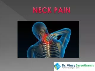
Neck
Neck. Thyroglossal duct cyst Failure of regression of the thyroglossal duct Prone to infection Require surgical excision Resection of midportion of hyoid and ligation of tract. Midline Neck. Thyroid nodules Common Greater incidence of malignancy in children


Neck
E N D
Presentation Transcript
Thyroglossal duct cyst • Failure of regression of the thyroglossal duct • Prone to infection • Require surgical excision • Resection of midportion of hyoid and ligation of tract Midline Neck
Thyroid nodules • Common • Greater incidence of malignancy in children • Twice as common in girls • Presentation • Midline cervical mass • Moves with thyroid • PE • Location • Associated lymphadenopathy Midline Neck
Thyroid nodules • Clinical findings unreliable • Imaging is rarely helpful • Multiple nodules suggestive of Hashimoto • FNA • Debated in pediatric population • Helpful if lesion benign • Surgical excision • Malignancy • Indeterminant • Benign lesions that cancer cannot be ruled out Midline Neck
Other • Midline branchial (cervical) cleft • Linear tract of epithelialized tissue in the anterior midline of the neck • Due to aberrant fusion of the branchial arches • Thymic cyst • Mediastinal lesions Midline Neck
A 3yo M returns to your clinic with a 4 ½ week history of a lateral neck mass. The mass is 4cm and firm. The Bartonella titers you ordered last week are negative. The child is otherwise healthy besides a recent URI and you suspect a mycobacterial infection. What is the treatment? A. Short course of antibiotics (1 week) B. Long course of antibiotics (4 weeks) C. Surgical excision D. Incision and drainage E. No treatment, this will resolve on its own Question 9
Most common • Benign reactive cervical lymphadenopathy • Nonspecific hyperplastic responses • URI or face/scalp infections • Characteristics • <2cm • Rubbery • Oval • Isolated • 2-10 y • Streptococcus pyogenes and Staphylococcus aureus • Spontaneous regression following resolution of infection Lateral Neck
Lymphadenitis • Bacterial infection of the node • Characteristics • Significant enlargement • Tenderness • Erythema • Suppuration • Treatment • Aggressive antibiotics • Surgical intervention if suppuration Lateral Neck
Chronic cervical lymphadenopathy • >4 weeks • DDx • Cat-scratch disease • Atypical mycobacterial infection • TB Lateral Neck
Cat-scratch disease • Common • Regional nodal enlargement 2-4 weeks following inoculation by dog or cat • Lymphadenopathy persists for several months • May require surgical drainage if suppurative • Diagnosis • Serologic testing • PCR of nodal tissue • Warthin-Starry stain on tissue specimen • Bartonella Lateral Neck
Mycobacterial infections • Various clinical presentation • Local adenopathy • Pulmonary infection • Disseminated disease • TB • Rare cervical or supraclavicularlymphadenopathy • Manifestation of significant intrathoracic disease • Treatment • Aggressive antimycobacterial therapy • Avoid surgery • Chronic draining sinus Lateral Neck
MAIC complex • Most common • Submandibular, submaxillary or preauricular lymph nodes • Characteristics • Large • Firm • Immobile • Nontender • May undergo spontaneous breakdown with abscess and sinus formation • Treatment • Complete resection Lateral Neck
Lymphoma • Characteristics • Painless cervical adenopathy • Absence of antecedent URI or cutaneous infection • Persistence of nodes beyond 6 weeks • Size >2cm • Firm • More common in Hodgkin • Incisional biopsy Lateral Neck
Branchial cleft anomalies • Second branchial cleft • Most common • Opening along the lower anterior border of SCM • Complete fistula • Drainage • Incomplete fistula • Cystic structure • Secondary infection is common • Treatment • Excision Lateral Neck
Fibromatosis coli • Fibrous dysplasia of the SCM • Mass in the lower neck • Tilting of the head and face to the side of the lesion • Older children may have hemifacialhypoplasia and asymmetry • Treatment • PT early • Prevents plagiocephaly and facial asymmetry • Surgery late Lateral Neck
Pectusexcavatum • “funnel chest” • Most common congenital chest wall deformity • Posterior angulation of the sternum toward the spine • 3M:1F • Deformity increases during childhood and adolescence • Treatment • Surgical repair • If symptomatic • Exercise intolerance, MVP, GER • If self-esteem problems Chest Wall
Pectuscarinatum • “pigeon chest” • Protrusion deformity • M>F • Usually asymptomatic • Consider Marfan • Aortic root abnormalities • Lens subluxation Chest Wall
Pectuscarinatum • Poland syndrome • Unilateral agenesis or dysplasia of the rib cage and chondral cartilages • Absence of pectoralis major and minor • Hand deformities • Breast and areolar defects Chest Wall
Most often lymphatic • Benign reactive lymphadenopathy • Most common mass • Cystic hygromas or lymphangiomas • May extend into mediastinum • Hiradenitis • Obstruction of sebaceous and sweat gland • Mass that may become superinfected and require surgical drainage Axilla
Mastitis • Common in infancy • May progress to abscess • Treatment • Aggressive antibiotic therapy • Warm compresses • Surgery – last resort • Permanent breast asymmetry and deformity Breast
Masses • Typically benign • Preadolescent (6-7y) • Firm mobile mass under one or both areolae • Precocious thelarche • Never biopsy • Adolescents • Fibroadenomas • 90% • Smooth and mobile • 1-2cm • Juvenile variant • Larger lesions with significant asymmetry • Low malignant potential • Treatment • Excision • Do not spontaneously resolve Breast
Masses • Fibrocystic disease • Older teens and young women • One or multiple firm, fixed and ill-defined msses • Hyperplasia of the fibrous parenchymal tissue • Variations throughout menstral cycle • Benign • Pubertal gynecomastia in males • Benign overgrowth of glandular tissue • Early puberty • Surgical intervention if psychosocial problems Breast
Masses • Phyllodes tumors • Rare fibroepithelial tumors • Benign with aggressive local behavior • May lead to malignancy and distant metastases • Surgical evaluation early • Nipple discharge • Purulence • Infection • Green or brown • Cyst • Bloody • Intraductalpapilloma in children • Cancer in adults • Galactorrhea • Endogenous hormones from tumor or pregnancy Breast
Mass evaluation • History and Family History • Mammography • Limited due to dense tissue • US • Good for cystic lesions • Needle aspiration • MRI • Chest wall involvement • Most masses can be serially monitored • Excisional biopsy may be indicated for a small group of postpubertal girls with lesions increasing in size. Breast
Omphalocele • Embryonic extrusion of the developing midgut while abdominal wall expands • Defect in the abdominal wall • Covered by a sac • Outer amniotic • Inner peritoneal • Umbilical cord insertion • Wide range of sizes • Coexisting anomalies • 30-50% • Heart • Sternum • Diaphragm • Bladder • Chromosomal Abdominal Wall
Gastroschisis • Defect of the right lateral abdominal wall • May be due to vascular accident leading to disruption of abdominal wall • Defect is usually small • May have large amount of bowel extruded • Bowel in contact with amniotic fluid • Intense inflammatory response or “peel” • Peel may alter bowel motility post op • 7-10% associated conditions • Intestinal atresias • Volvulus, malrotation or incarceration Abdominal Wall
Management • Goal • Safe primary closure • Staged closure • Prosthetic Silastic silo with daily reductions • Prosthetic material • Complications • Abdominal compartment syndrome • Pulmonary embarrassment • Renal insufficiency • Intestinal ischemia or NEC • Timing • Gastroschisis – emergent • Omphalocele – more elective Abdominal Wall
Umbilical hernia • Most common condition of the abdominal wall • Failed closure of the fascial ring • Usually closes in 2-3 years • Strong familial and racial predilection • AA • Treatment • Repair • Delay until age 5 • >2cm • “elephant’s trunk” • Incarceration Umbilicus
Granulomas • Polypoid mucosal-appearing lesion at the base of umbilicus • Residual tissue at the base of the cord • Treatment • Topical • Alcohol • Silver nitrate sticks Umbilicus
Hernias • Presentation • Bulges in the groin and scrotum or labial majora • Increase with valsalva • Reduces spontaneously or with pressure • Complications • Incarceration • Unable to reduce • 30% • Treatment • Prompt repair • No spontaneous resolution Inguinal Disorders
Acute scrotal inflammation • Incarcerated hernia • Torsion of the testicle • Torsion of the appendix testis • Testicular trauma • Epididymo-orchitis • EMERGENCY Inguinal Disorders
Testicular torsion • Acutely tender testicle • Retracted toward inguinal region • Transverse lie • Immediate surgery • Torsion of appendix testis • Blue dot sign • Does not require surgery Inguinal Disorders
At what age should this patient be referred to surgery if the condition has not resolved? A. 1-3 months B. 3-6 months C. 6-9 months D. 12-18 months E. 2 years Question 10
Hydroceles • Common in infancy • Diminish during childhood • Characteristics • Scrotal swellings • Diurnal variation • Transillumination • Birth • Noncommunicating • Resolve by 1 year • Surgery • Communicating • Persistent >12-18 months of age • Persistent after infection or trauma Inguinal Disorders
Imperforate anus • Low • Passage of the rectum through the levatorani • Fistulous tract to perineal region ending • Center of a ridge of tissue • Bucket handle deformity • Anterior to the structures as a perineal fistula • Favorable prognosis • Passes through levatorani Anus and Rectum
Imperfortate anus • High • No visual fistula • No levatorani • Fistula to • Prostatic urethra • Bulbar urethra • Bladder neck • Hymen • Meconium passed with urine or transvaginally • Management • Perinealanoplasty • One or multiple stages involving colostomy • May change in future • Prognosis guarded due to other anomalies Anus and Rectum
Rectal prolapse • Uncommon • Most often idiopathic • Peak at 2y • Precipitated by • Diarrheal illness • Toilet training • Severe constipation • Management • Resolves spontaneously • Dietary or medical manipulations for constipation Anus and Rectum
Rectal prolapse • Nonidiopathic • Spina bifida • Other spinal cord abnormalities • Chronic hookworm infestation • Rectal polyps • CF • Management • Circumferential submucosal injections with concentrated dextrose • Sclerosant Anus and Rectum
Anorectal abscess • Common 6-10 months • Infection of submucosal crypt glands • Complications • Recurrent episodes lead to fistulas or chronically draining sinuses • Crohn, CGD, Immunodeficiency • Management • I & D if fluctuant • Warm soaks • Sitz baths • Anal fistulectomy Anus and Rectum