Download
1 / 20
200 likes | 449 Vues
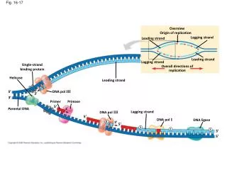
Overview. Origin of replication. Lagging strand. Leading strand. Fig. 16-17. Leading strand. Lagging strand. Single-strand binding protein. Overall directions of replication. Helicase. Leading strand. 5 . DNA pol III. 3 . 3 . Primer. Primase. 5 . 3 . Parental DNA.

E N D
Overview Origin of replication Lagging strand Leading strand Fig. 16-17 Leading strand Lagging strand Single-strand binding protein Overall directions of replication Helicase Leading strand 5 DNA pol III 3 3 Primer Primase 5 3 Parental DNA Lagging strand DNA pol III 5 DNA pol I DNA ligase 4 3 5 3 2 1 3 5
Figure 16.18 DNA pol III Leading strand Parental DNA 5 5 3 3 3 5 3 5 Connectingprotein Helicase Laggingstrandtemplate 3 5 DNA pol III Lagging strand 3 5
Cool animation of DNA replication (and other stuff) http://www.ted.com/talks/drew_berry_animations_of_unseeable_biology.html
How do we know all this? • That the lagging strand is made in fragments • That DNA ligase joins these fragments together
1 2 TECHNIQUE Powersource Gel electrophoresis Mixture ofDNA mol-ecules ofdifferentsizes Anode Cathode Wells Gel Powersource Longermolecules Shortermolecules
Where do mutations come from? DNA Pol III
How often do mutations occur? ~1 “error” for every 1010bases replicated E.Coli genome: 4.6 x 10^6 b.p. H. Sapiens genome (diploid): 6 x 10^9 b.p. DNA pol III adds the wrong base every 105 bases Extra fidelity comes from: 1. “Proofreading” by DNA pol III (and pol I) 2. Mismatch repair pathway
How often do mutations occur? ~1 “error” for every 1010bases replicated E.Coli genome: 4.6 x 10^6 b.p. H. Sapiens genome (diploid): 6 x 10^9 b.p. DNA pol III adds the wrong base every 105 bases
How often do mutations occur? ~1 “error” for every 1010bases replicated E.Coli genome: 4.6 x 10^6 b.p. H. Sapiens genome (diploid): 6 x 10^9 b.p. DNA pol III adds the wrong base every 105 bases Extra fidelity comes from: 1. “Proofreading” by DNA pol III (and pol I) 2. Mismatch repair pathway Chemically damaged DNA can lead to much higher rates of mutation
Example of damaged DNA: thymine dimer caused by UV radiation Nuclease Fig. 16-18 DNA polymerase DNA ligase Nucleotide Excision Repair pathway has removed and replaced damaged bases
Example of damaged DNA: thymine dimer caused by UV radiation Nuclease Fig. 16-18 Nucleotide Excision Repair pathway removes and replaces damaged bases DNA polymerase DNA ligase
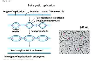
Eukaryotic replication Origin of replication Double-stranded DNA molecule Parental (template) strand Daughter (new) strand Fig. 16-12b 0.25 µm Replication fork Bubble Two daughter DNA molecules (b) Origins of replication in eukaryotes
5 Ends of parental DNA strands Leading strand Lagging strand 3 Last fragment Previous fragment RNA primer Lagging strand 5 Fig. 16-19 3 Parental strand Removal of primers and replacement with DNA where a 3 end is available 5 3 Second round of replication 5 New leading strand 3 5 New lagging strand 3 Further rounds of replication Shorter and shorter daughter molecules
5 Ends of parental DNA strands Leading strand Lagging strand 3 Last fragment Previous fragment RNA primer Lagging strand 5 Fig. 16-19 3 Parental strand Removal of primers and replacement with DNA where a 3 end is available 5 3 Second round of replication 5 New leading strand 3 5 New lagging strand 3 Further rounds of replication Shorter and shorter daughter molecules
Staining of telomeres Florescence In Situ Hybridization (FISH) Fig. 16-20 1 µm “probe” = (5’-CTAACC-3’)100
5 end Hydrogen bond 3 end 1 nm 3.4 nm Fig. 16-7a 3 end 0.34 nm 5 end (a) Key features of DNA structure (b) Partial chemical structure
Fig. 16-21a Nucleosome (10 nm in diameter) DNA double helix (2 nm in diameter) H1 Histone tail Histones DNA, the double helix Histones Nucleosomes, or “beads on a string” (10-nm fiber)
Chromatid (700 nm) 30-nm fiber Fig. 16-21b Loops Scaffold 300-nm fiber Replicated chromosome (1,400 nm) 30-nm fiber Looped domains (300-nm fiber) Metaphase chromosome