Download
1 / 27
270 likes | 639 Vues
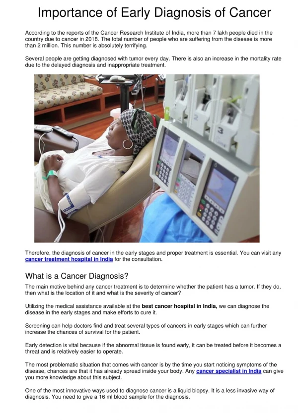
C41 Importance of Early Detection. Objectives. State thesis Identify breast anatomy Describe types of breast tissue Define breast cancer Explain types of breast cancer Identify risks and signs of breast cancer Determine diagnosis process Assess treatment options and prognosis. Thesis.

E N D
Objectives • State thesis • Identify breast anatomy • Describe types of breast tissue • Define breast cancer • Explain types of breast cancer • Identify risks and signs of breast cancer • Determine diagnosis process • Assess treatment options and prognosis
Thesis Early detection is key in providing an optimistic prognosis for women diagnosed with breast cancer
Breast Anatomy The breast tissue begins under the clavicle to below breast, axilla to sternum and includes the pectoral muscle https://nbcf.org.au/about-national-breast-cancer-foundation/about-breast-cancer/what-you-need-to-know/breast-anatomy-cancer-starts/
Breast Tissue Anatomy • The breast tissue is made up of fatty tissue, lobules, lobes and ducts • Lobules: Gland that produces milk in women who are lactating • Lobes: Made up of numerous lobules • Both the lobules and the lobes connect directly to the ducts • Ducts: Tubes that carry milk to the nipple during lactation https://moxxly.com/blogs/modern-mom-guides/nipples-101
Types of Breast Tissue There are four types of breast tissue: fatty, fibro glandular, heterogeneously dense, extremely dense Dense breast tissue is the most difficult to evaluate and make an accurate diagnosis https://www.mayoclinic.org/tests-procedures/mammogram/in-depth/dense-breast-tissue/art-2012396 https://www.itnonline.com/article/mammographic-breast-density-%E2%80%94-what-it-means
What Is Breast Cancer? Type of cancer that occurs when the cells in the breast begin to grow in an abundanceand form a tumor These cells can be found in the ducts, lobes or the glands Breast cancer is the number one cause of cancer in women: 1 in 8 women develop breast cancer
Breast Cancer Types There are many types of breast cancer; however can be categorized into two main types: Noninvasive (in situ) and Invasive Noninvasive: Often referred to as In Situ (in its original place). The cells do not break away and spread to any other healthy tissue • Ductal Carcinoma In Situ (DCIS): Abnormal cells are found in the lining of breast milk ducts. These abnormal cells have not spread or invaded beyond the walls of the ducts into surrounding breast tissue • Lobular Carcinoma In Situ (LCIS):Confined to the milk-producing glands(lobules) of the breast. Tumors classified as LCIS are made up of small cells that do not leave the lobules. Invasive: Any cancerous cell that invades normal (healthy) tissue is deemed invasive. • Invasive Ductal Carcinoma (IDC): Originate in the milk ducts of the breast and travel to surrounding tissue. • Invasive Lobular Carcinoma (ILC): Begin in the lobules of the breast. The cells in the lobules disperse and invade surrounding tissue.
Risk Factors • Gender:1 in 8 women will develop breast cancer • Age:Risk increases after 45 years old • Ethnicity: Non-Hispanic whites have the highest risk • Family history: Maternal and Paternal • Genetics: Inheriting the gene mutation (BRCA1/BRCA2) • Previous Radiation: Specific to breast area • Other risk factors include: Smoking, being overweight, consuming more than 2-3 alcoholic beverages per day, not bearing children, and not breast feeding
Signs Of Breast Cancer https://mymodernmet.com/know-your-lemons-update/
Diagnosing Breast Cancer • Selfand clinical breast exam • A self breast exam is the first step. Consists of using the fingertips to press against the entire breast tissue to feel for any lumps or suspicious changes in the breast. These should be completed monthly. • A clinical breast exam should be completed from the patients primary care provider during the annual physical. • Imaging modalities: • Mammography • Tomosnythesis • Magnetic Resonance Imaging (MRI) • Ultrasound • Breast Biopsies
Mammography • Gold standard of imaging for breast cancer • During a mammogram, the patient’s breast is placed between two flexible, radiolucent paddles that compress and flatten the breast tissue. This compression allows for the layers of the breast tissue to be spread apart, allowing for less overlapping of the tissue in the breast. • Limitations to mammography: • Low detection rate for those women with dense breast • High false positive recall rate (results that show a person has a disease when they do not) https://www.ncbi.nlm.nih.gov/books/NBK65716/figure/CDR0000062970__386/
Mammography-Images https://nutreaunnino.com/craniocaudal-mammogram-anatomy http://missbelmonte.weebly.com/mammograms.html
Tomosynthesis • New screening tool that features pseudo 3D digital imaging; uses a low-dose x-ray system • Obtains multiple thin, high-resolution 2D slices and reconstructs them into 3D images • Provides better imaging for those women who have dense breast tissue; requires more angles. This results in an increase in accuracy of size, shape, localization of any tumors that may be detected • Uses increased compression allowing: • Less blurring on the image from patient motion • Tissue to be spread out further, which causes the tissue to become thinner • Thinner tissue will provide smaller abnormalities to be better detected on the images • Thinner tissue also means less dose to the patient • With smaller abnormalities being detected it provides earlier diagnosis of breast cancer, which can lead to more treatment options and a more positive prognosis
Mammography vs. Tomosynthesis Breast Cancer in 2D (mammography) vs. 3D (Tomosynthesis) https://www.healthdatamanagement.com/news/study-finds-radiologists-easily-transition-to-3d-mammography https://www.healthcarefinancenews.com/news/3-d-mammography-bests-digital-screenings-costs-diagnosis
MRI • MRI is a nonionizing imaging tool that uses strong magnets and radio waves that interact with the body to produce detailed images of organs and soft tissues in the body • Used to aide in detecting the presence, stage and/or type of breast cancer • If cancer is detected, MRI will assist in measuring the size and shape of the detected lesion https://breast-cancer.ca/mrifacts/
Ultrasound • Ultrasound uses sound waves to help differentiate between a solid mass and a fluid-filled cysts in the breast • A fluid filled cyst detects different sound wave patterns that a solid structure • Ultrasound will not specify whether the tumor is cancerous or benign • Used for patients who have suspicious findings on a mammography or MRI. http://www.startradiology.com/the-basics/ultrasound-technique/ https://blog.insightdatascience.com/automating-breast-cancer-detection-with-deep-learning-d8b49da17950
Breast Biopsies • Performed when other imaging detects suspicious findings • Aide in diagnosing abnormalities in the breast tissue • Samples are obtained and sent to lab for testing • Lab reports assist in determining a patient’s treatment plan • MRI, ultrasound and x-radiation imaging guide the retrieval of fluid or small specimen https://www.mayoclinic.org/tests-procedures/breast-biopsy/about/pac-20384812 Ultrasound guided biopsy
Treatment Options • Once diagnosed, a patient may endure one or more of the following: • Surgical • Radiation therapy • Chemotherapy
Surgery Lumpectomy Mastectomy Surgical removal of the entire breast and lymph nodes to eliminate any and all tissue that may contain breast cancer May have a single (one breast) or a double (both breast) mastectomy • Surgical removal of a lump that presents as cancerous inside the breast tissue • Less invasive than mastectomy Both techniques are methods to reduce the chance of cancer spreading or returning to the same area
Radiation Therapy • Uses ionizing radiation to direct all radiation dose solely to the cancerous cells and destroy them • Patients may receive radiation 5 days in a row, lasting up to 6-8 weeks • This form of treatment can be completed before or after surgery • Before surgery: Minimizes the tumor before removal (less tissue has to be removed) • After surgery: Targets the area removed in order to ensure all cancerous tissue was fully removed https://www.mayoclinic.org/tests-procedures/radiation-therapy/about/pac-20385162 Radiation therapy machine
Chemotherapy • Type of treatment that uses chemical substances that are administered intravenously over a course of time in cycles to kill the cancerous cells throughout the entire body • Usually done for patients with metastatic breast cancer • Administered either before or after surgery: • Before Surgery: Reduces the size of the tumor which results in less tissue being removed during surgery • After Surgery: Eliminates any cancerous cells that may still be present and/or to reduce the chances of the cancer returning • Most intensive treatment option • Because it is treating the whole body • Chemicals used causes lots of side effects such as: nausea, vomiting, appetite changes, severe constipation, and hair loss
Early Detection is Key • In order to avoid intensive treatment, such as chemotherapy, the focus of early detection is vital • Qualities of early detection: • Women being aware of their risk factors, so they know when to start their initial mammogram. This aides in detection of smaller lesions • Being aware of their signs and to take actions early • Obtaining a mammogram with tomosynthesis software increases early detection • When a patient is diagnosed early, they are provided with more treatment options
Follow-up Care • Monthly visits with the patient’s primary care provider to evaluate the patients body physically and mentally • Mammograms: follow up screening every six months or annually, dependent on how long a patient has gone without breast cancer
Prognosis • Due to earlier detection, 90% of women who have been diagnosed with breast cancer, survive for at least five years after initial diagnosis • Currently, there are 3.1 million (women) breast cancer survivors in the United States • It is estimated that 40,920 women succumbed to the disease in 2017
Conclusion • Breast cancer is the number one cause of cancer in women • Women who get self breast exams, annual screenings with tomosynthesis provide early diagnosis • Once diagnosed, women are faced with treatment options that include: • Surgery • Radiation Therapy • Chemotherapy • Early detection offers an optimistic prognosis of 90% survival of those diagnosed
References • October Is Breast Cancer Awareness Month: 1 in 8 Women Will Develop Breast Cancer In Her Lifetime. Racine Journal Times. October 10, 2018:Section B. • Breast Cancer. American Cancer Society. https://www.cancer.org/cancer/breast-cancer.html. Published 2018. Accessed October 15, 2018. • Eisenberg RL, Johnson NM. Mammography. Comprehensive Radiographic Pathology. 6th ed. St. Louis, MO:Mosby Inc.; 2016:746-759. • Search Results. Mayo Clinic. https://www.mayoclinic.org/search/search-results?q=breast cancer. Published 2018. Accessed October 8, 2018. • Coop P, et al, Tomosynthesis as a screening tool for breast cancer: A systematic review. Radiography.2016;22(3):190-195. doi:http://dx.doi.org/10.1016/j.radi. 2016.03.002. • Breast Cancer Diagnosis, Treatments & Therapies | CTCA. CancerCenter.com. https://www.cancercenter.com/breast-cancer/diagnostics-and-treatments/. Published January 1, 1AD. Accessed October 8, 2018. • White N. For October, here are 31 facts (one a day) about breast cancer. Breast cancer surgery, treatment, screening | City of Hope Cancer Hospital. https://www.cityofhope.org/blog/31-facts-about-breast-cancer. Published October 24, 2017. Accessed November 18, 2018. • Radiological Society of North America, RSNA, American College of Radiology. Breast Tomosynthesis. RadiologyInfo.org. https://www.radiologyinfo.org/en/info.cfm?pg=tomosynthesis. Published 2018. Accessed November 10, 2018. • Who Gets Chemotherapy as Treatment? Breastcancer.org. https://www.breastcancer.org/treatment/chemotherapy/who_gets_it. Published 2018. Accessed November 11, 2018.